
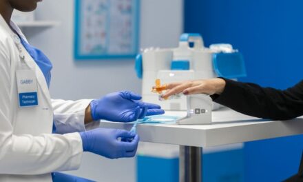
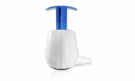
Akoya Biosciences, Inc., The Spatial Biology Company, has announced the commercial availability of the PhenoCycler-Fusion system for high-speed imaging of whole slides, at single-cell and sub-cellular resolution.
The PhenoCycler-Fusion platform fuses the strengths of the company’s automated, ultrahigh multiplex cycling platform, PhenoCycler (previously branded as CODEX), and its high-speed imaging platform, PhenoImager (previously branded as Phenoptics), into an end-to-end, integrated workflow. The increased speed of its workflow enables researchers to image multiple biomarkers and millions of cells across whole slides in just minutes, providing an unbiased view of how cells spatially organize and interact to influence disease outcomes.
A benefit of the system is its tunable workflow that offers researchers the flexibility to run large panels for hypothesis-free biomarker discovery and focused panels for high-throughput biomarker validation, all on a single system. This capability simplifies the translation of spatial biomarker signatures onto the PhenoImager HT, a high-throughput platform that can process 300+ samples per week, according to the company.
“The PhenoCycler-Fusion delivers on our vision of providing an integrated solution to scale up spatial biology studies that require larger panels, higher sample throughput and an unbiased interrogation of every cell from a tissue sample.” says Brian McKelligon, chief executive officer of Akoya Biosciences.
In addition to its well-established protein biomarker assays, the system will also be enabled with RNA detection to provide a deeper, comprehensive view into tissue biology. To this end, Akoya recently announced a partnership with Bio-Techne, to automate RNAScope assays, a highly cited and proven standard in RNA imaging, on the PhenoCycler-Fusion system. This multiomic workflow will also complement a novel spatial transcriptomics chemistry, currently being developed by Akoya, which will also be compatible with PhenoCycler-Fusion.
“Spatial discovery is all about studying the rich diversity of cell types, cellular neighborhoods and their interactions, across the entirety of a tissue section.” says Garry Nolan, Ph.D., the Rachford and Carlota A. Harris Professor in the Department of Microbiology and Immunology at Stanford University School of Medicine and a leading pioneer in the field of spatial biology. “This can happen on a much larger scale if we can image whole slides with a high-speed system like PhenoCycler-Fusion. The throughput, flexibility, and compatibility with archived slides are some of the reasons we decided to adopt the platform in our lab.”
The PhenoCycler-Fusion offers the following features:
- Single-cell and sub-cellular resolution, at a standard long established by Akoya’s spatial phenotyping systems
- Ultrahigh plex detection of 100+ markers for deep spatial phenotyping
- Rapid, unbiased mapping of every cell across entire tissue sections, including archived FFPE (formalin-fixed, paraffin-embedded) slides
- High-speed imaging that enables the processing of 100+ whole slides per week.
- Multiomic detection of protein and RNA biomarkers
Akoya has also rebranded its existing products—CODEX is now PhenoCycler, the Phenoptics workflow is renamed as PhenoImager, and the Vectra Polaris instrument is the PhenoImager HT.
“We’ve been using the CODEX platform for studying the spatial interactions of tumor and immune cells and how they behave in response to immunotherapy” says Robert Schreiber, Ph.D., Professor of Pathology and Immunology, Washington University, St. Louis and an expert in immuno-oncology. “With PhenoCycler-Fusion we can now scale up to higher throughput studies with large cohorts of samples and image entire slides in a rapid timeframe.”
For more information about PhenoCycler-Fusion, please visit Akoya Biosciences.

