 Digital Imaging and Analysis System
Supports all nuclear and membrane stains
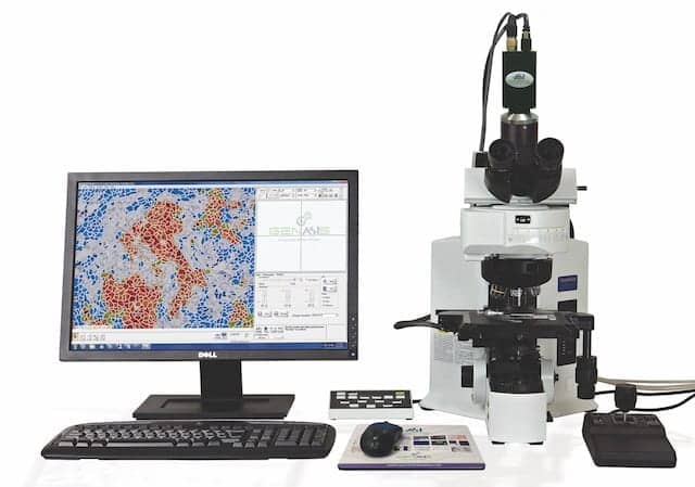
Genasis HiPath from Applied Spectral Imaging, Carlsbad, Calif, is a digital imaging and analysis system for immunohistochemistry tissue samples. Designed to provide accuracy and standardization of results, the system aims to bring new capabilities to the pathologist’s microscope. The complete digital solution is available at a fraction of the cost of whole-slide imaging systems, while maintaining existing workflows and methodologies. HiPath is FDA cleared for the analysis of ER, HER2/neu, Ki-67, and PR status, and supports all nuclear and membrane stains, including those for PD-L1.
Applied Spectral Imaging
(800) 611-3466; www.spectral-imaging.com
 On-Demand Digital Pathology System
Offers turnkey solution
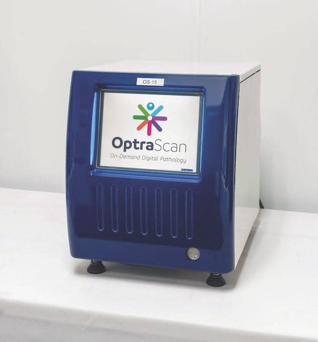
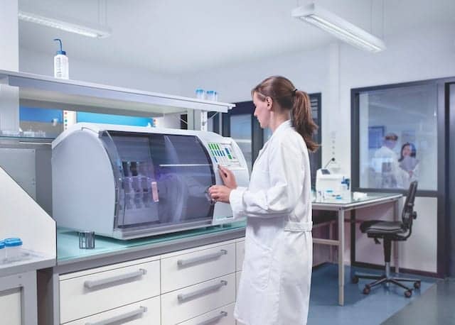
OptraScan from OptraScan Inc, Sunnyvale, Calif, is an on-demand digital pathology system developed to provide a turnkey solution for low- and high-throughput users. OptraScan serves as a tool for the acquisition of whole-slide imaging (WSI) during the transition from conventional microscopy to digital pathology, enabling viewing, sharing, analysis, and management of digital slides and associated metadata. The system includes the small-footprint OptraScan WSI scanner (15- or 120-slide brightfield, frozen sections, fluorescent), the ImagePath integrated image viewer and management system, the TelePath telepathology system, the OptraAssays image analysis and computer-aided region detection system (CARDS), and 10 TB of complimentary cloud storage. For research use only.
OptraScan
(408) 524-5300; www.optrascan.com
 Quantitative Image Analysis System
Enables multiplexing
The Onoctopix from Visiopharm Corp, Copenhagen, Denmark, is a quantitative image analysis system for cancer research and diagnostics. Onoctopix features Tissuealign, a module to align and subsequently analyze digitized images of serial tissue sections. Tissuealign provides high-precision alignment of any number of scanned tissue sections independently of tissue and image modality. A key application for diagnostics is automated tumor cell detection using such tumor markers as cytokeratin, hematoxylin and eosin stained tissue sections, and melanoma antigen. The system enables multiplexing and colocalization analysis, revealing more information about the tumor microenvironment than ever before. For research use only outside Europe.
Visiopharm
(877) 843-5268; www.visiopharm.com
 Software Suite
Includes universal viewer
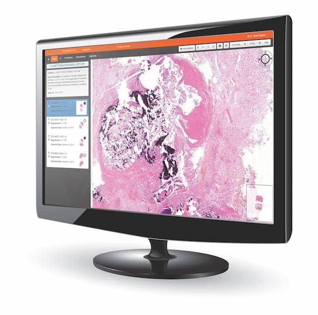
The DP3 software suite from Corista, Concord, Mass, is helping the nation’s leading hospitals and research centers harness the power of digital pathology to streamline their practices while energizing telepathology, tumor boards, peer-reviewed quality assurance, and investigator-initiated research efforts. DP3 is a vendor-agnostic digital pathology platform that offers a universal viewer to support synchronous, remote consultation among any number of physicians, anywhere in the world. DP3 delivers an extensive array of workflow, analytical, and collaborative tools while seamlessly integrating whole-slide, gross, and static images with data from any laboratory information system and electronic health record.
Corista
(978) 287-6188; www.corista.com
 Integrated Diagnostics Platform
Aims to improve clinical decisionmaking
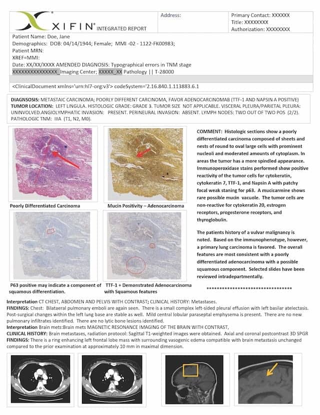
ProNet from Xifin Inc, San Diego, expands the use of patient data in clinical environments to improve decisionmaking and support cost-effective, patient-centered care. ProNet enables hospitals and health systems to integrate and exchange diagnostic images from different modalities along with other clinical and patient encounter information into a live, shared clinical workflow. Clinicians use the cloud-based information exchange capabilities to collaborate on complex cases, expedite treatment decisions, and increase efficiencies in geographically dispersed environments. Xifin enables integration with electronic health records via web services and HL7 interfaces.
Xifin
(858) 793-5700; www.xifin.com
 Digital Pathology Software
Provides data management and image analysis
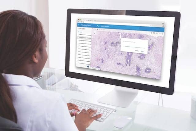
Proscia Inc, Baltimore, provides pathologists with an intuitive and secure platform to access, analyze, and share whole-slide images anywhere in the world. Part of the modern pathology workflow, Proscia’s vendor-neutral technology integrates with any whole-slide image scanner and information management system. Vendor lock-in and information technology are not required to implement the software. Users can remotely access data and slides, collaborate on tumor boards and second-opinion consultations, and leverage enhanced quantitative image analysis tools and algorithms to identify patterns and pixels unseen by the human eye.
Proscia
(877) 255-1341; www.proscia.com
 Digital Pathology Solution
Simplifies rad-path collaboration
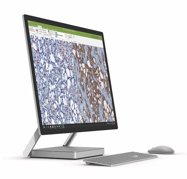
Sectra, Linköping, Sweden, provides a complete and vendor-neutral solution for primary diagnostics in pathology. The review workstation provides tools to perform daily tasks in a digital system and to reduce pain points associated with time-consuming manual workflows. It provides full case overview, including images from different scanners, macro cameras, and other image systems, as well as patient information from electronic medical records and laboratory information systems, in the same workstation. With support for all kinds of medical imaging, the system also enables efficient tumor boards and simplifies rad-path collaboration.
Sectra
(203) 925-0899; www.sectra.com
 Whole-Slide Imaging System
Features advanced software tools
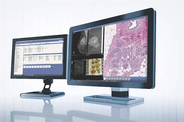
The IntelliSite pathology solution from Philips Medical Systems BV, Amsterdam, is a whole-slide imaging system that allows for review and interpretation of digital pathology slides prepared from biopsied tissue. The IntelliSite system comprises an ultrafast pathology slide scanner, an image management system, and a display, to automate the creation, viewing, and management of digital pathology images. The system is complemented by advanced software tools to manage the scanning, storage, presentation, review, and sharing of information. The system’s recent de novo premarket notification (510(k)) clearance marks the first time that FDA has permitted the marketing of a digital pathology solution for primary diagnostic use in the United States.
Philips Medical Systems
(800) 229-6417; www.usa.philips.com
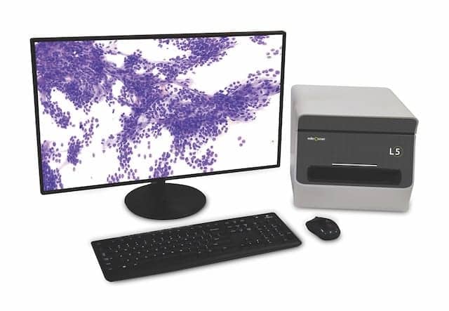
Robotic Microscope
Supports plug-and-play remote control
Mikroscan Technologies, Carlsbad, Calif, has released the L5 telemicroscopy system, which supports remote-controlled microscopy. Its live-only functionality enables efficient telemicroscopy in a small footprint. Users interact with the actual glass slide from a distance, enabling them to provide on-demand microscopy whenever and wherever it is needed. The system features five objectives ranging in magnification from 2x to 40x, with a 60x option. Complete remote control of the X, Y, and Z axes, plus objective magnification selection and contrast adjustments, are included to further enhance the solution’s live capabilities.
Mikroscan
(844) 213-3452; www.mikroscan.com
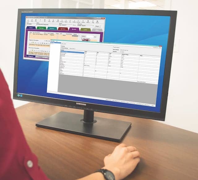 Clinical Results Management Module
Produces integrated or standalone reports
NovoPath Inc, Princeton, NJ, offers a clinical results management module supporting the growing demand for integration of anatomic pathology results with clinical results. Using the module, laboratories have the flexibility to produce integrated anatomic pathology reports that contain clinical pathology results, a standalone anatomic pathology report, or just a clinical pathology report. The clinical information can also be quickly and accurately imported into NovoPath from other software systems or instruments and displayed in tabular form for quick referencing, workflow enhancement, and patient safety.
NovoPath
(877) 668-6123; www.novopath.com
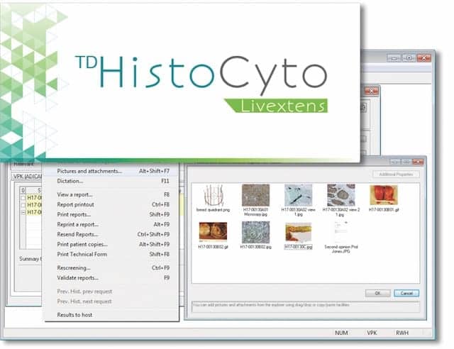 Laboratory Information System
Facilitates paperless communication
TDHistoCyto from Technidata, Montbonnot, France, is a dedicated software suited to the specific needs of anatomic pathology laboratories. The laboratory information system seeks to improve lab processes and efficiency, enhance patient safety and traceability, and adopt paperless communication. The software supports such practices as image management, adjunctive testing on samples obtained by liquid-based cytology, and connections with cassette and slide printers, and with digital pathology and immunohistochemistry instruments. The system is designed to reduce processing and diagnosis times, monitor turnaround times and activity levels, maximize traceability, and lower the risk of errors for faster, more reliable diagnosis.
Technidata
(514) 270-7777; www.technidata-web.com
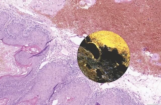 Whole-Slide Alignment Software
Includes import wizard
MicroDimensions, Munich, has launched a new version of its high-precision whole-slide alignment software, Slidematch. Users can now align digital whole-slide images from consecutive sections in brightfield and fluorescence mode, improving suitability for applications such as the evaluation of HER2 status of cancer patients. An import wizard allows users to load images in formats from different vendors and in different resolutions in the same batch. The images can be stored as an aligned image series in .svs, .tif, or .ims formats for viewing or automated image analysis.
MicroDimensions
www.micro-dimensions.com
