Summary: A new automated device can diagnose glioblastoma, a highly aggressive brain cancer, in under an hour using a novel electrokinetic biochip that detects active Epidermal Growth Factor Receptors (EGFRs) from blood samples.
Takeaways:
- Rapid Diagnosis: The new device can diagnose glioblastoma in less than an hour, significantly reducing the time needed for detection compared to traditional methods.
- High Sensitivity and Selectivity: The biochip’s electrokinetic sensor enhances sensitivity and selectivity by detecting active EGFRs on extracellular vesicles without requiring blood sample pretreatment, minimizing interference from other particles.
- Broad Applicability: Although initially developed for glioblastoma, the technology can be adapted to detect other biomarkers, potentially enabling early diagnosis of various diseases, including pancreatic cancer and cardiovascular disorders.
Researchers at the University of Notre Dame have developed a novel, automated device capable of diagnosing glioblastoma, a fast-growing and incurable brain cancer, in less than an hour. The average glioblastoma patient survives 12-18 months after diagnosis.
Detecting Active Epidermal Growth Factor Receptors
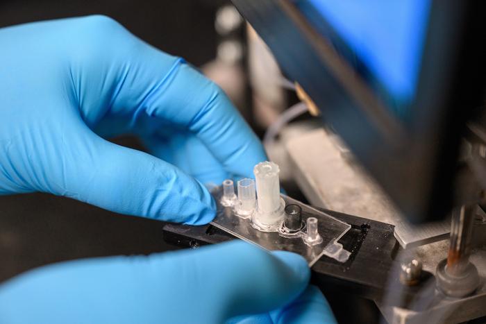
The crux of the diagnostic is a biochip that uses electrokinetic technology to detect biomarkers, or active Epidermal Growth Factor Receptors (EGFRs), which are overexpressed in certain cancers such as glioblastoma and found in extracellular vesicles.
“Extracellular vesicles or exosomes are unique nanoparticles secreted by cells. They are big — 10 to 50 times bigger than a molecule — and they have a weak charge. Our technology was specifically designed for these nanoparticles, using their features to our advantage,” says Hsueh-Chia Chang, the Bayer Professor of Chemical and Biomolecular Engineering at Notre Dame and lead author of the study about the diagnostic published in Communications Biology.
The challenge for researchers was two-fold: to develop a process that could distinguish between active and non-active EGFRs, and create a diagnostic technology that was sensitive yet selective in detecting active EGFRs on extracellular vesicles from blood samples.
Creation of a New Biochip
To do this, researchers created a biochip that uses an inexpensive, electrokinetic sensor about the size of a ball in a ballpoint pen. Due to the size of the extracellular vesicles, antibodies on the sensor can form multiple bonds to the same extracellular vesicle. This method significantly enhances the sensitivity and selectivity of the diagnostic.
Then synthetic silica nanoparticles “report” the presence of active EGFRs on the captured extracellular vesicles, while bringing a high negative charge. When extracellular vesicles with active EGFRs are present, a voltage shift can be seen, indicating the presence of glioblastoma in the patient.
This charge-sensing strategy minimizes interference common in current sensor technologies that use electrochemical reactions or fluorescence.
“Our electrokinetic sensor allows us to do things other diagnostics cannot,” says Satyajyoti Senapati, a research associate professor of chemical and biomolecular engineering at Notre Dame and co-author of the study. “We can directly load blood without any pretreatment to isolate the extracellular vesicles because our sensor is not affected by other particles or molecules. It shows low noise and makes ours more sensitive for disease detection than other technologies.”
Components of the New Diagnostic Device
In total, the device includes three parts: an automation interface, a prototype of a portable machine that administers materials to run the test and the biochip. Each test requires a new biochip, but the automation interface and prototype are reusable.
Running one test takes under an hour, requiring only 100 microliters of blood. Each biochip costs less than $2 in materials to manufacture.
Adaptable to Other Biological Nanoparticles
Although this diagnostic device was developed for glioblastoma, the researchers say it can be adapted for other types of biological nanoparticles. This opens up the possibility for the technology to detect a number of different biomarkers for other diseases. Chang says the team is exploring the technology for diagnosing pancreatic cancer and potentially other disorders such as cardiovascular disease, dementia, and epilepsy.
“Our technique is not specific to glioblastoma, but it was particularly appropriate to start with it because of how deadly it is and the lack of early screening tests available,” Chang says. “Our hope is that if early detection is more feasible, then there is an increased chance of survival.”
Blood samples for testing the device were provided by the Centre for Research in Brain Cancer at the Olivia Newton-John Cancer Research Institute in Melbourne, Australia.
In addition to Chang and Senapati, other collaborators include former postdocs at Notre Dame Nalin Maniya and Sonu Kumar; Jeffrey Franklin, James Higginbotham and Robert Coffey from Vanderbilt University; and Andrew Scott and Hui Gan from the Olivia Newton-John Cancer Research Institute and La Trobe University. The study was funded by the National Institutes of Health Common Fund.
Chang and Senapati are affiliated with Notre Dame’s Berthiaume Institute for Precision Health, the Harper Cancer Research Institute, and NDnano.
Further reading: Biomarker for Brain Cancer Blood Test Identified
Featured image: The biochip is used to detect biomarkers for glioblastoma, a fast-growing brain cancer. Photo: Matt Cashore/University of Notre Dame


