This is a companion article to the feature, “Pathology Goes Digital.”
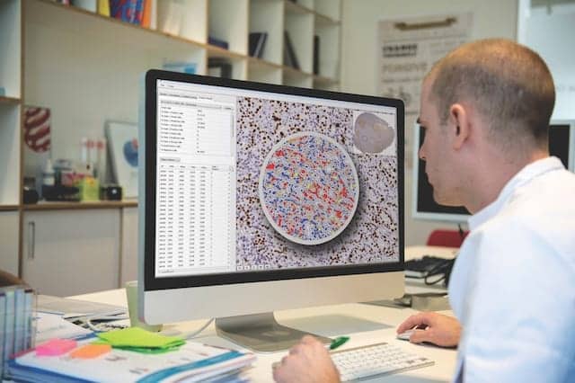
The Halo next-generation image analysis platform by PathXL offers precision histology analytics and purpose-built modules for developing quantitative results in metabolism, neuroscience, oncology, toxicological pathology, and other fields.
Important advantages of whole-slide imaging include its utility for advancing pathology education and training, and for supporting multidisciplinary teamwork in clinical settings.
Traditionally, education and training in pathology have been delivered using textbooks, glass slides, and conventional microscopy. Over the past decade, however, the number of Web-based pathology resources has expanded dramatically, with centralized pathology resources being delivered to many students simultaneously.
Whole-slide imaging technology enables glass slides to be scanned and viewed on a computer screen via dedicated software. Sometimes referred to as “virtual microscopy,’ this technology has created enormous opportunities in pathology education and training. Via a Web-based computer environment, virtual microscopy students are able to learn key histopathology skills—such as how to identify areas of diagnostic relevance from an entire slide—even when they are unable to be in the same room as either the slides or their instructor. Today, Philips offers an education solution, Tutor, coming from its acquisition of PathXL, that is a market-leading solution with more than 40,000 users worldwide.
Building on the basics of virtual microscopy to broaden its applications in educational settings, new human-computer interfaces using natural-touch technology are being developed to enhance the manipulation of digitized slides. Several major initiatives are under way to adapt virtual microscopy to online competency and diagnostic decision analysis, with important implications for the future of professional accreditation and recertification in pathology.
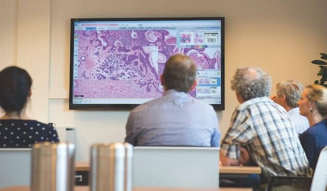
With whole-slide images displayed by the IntelliSite pathologist suite, a tumor board discusses a cancer case.
And researchers are also using virtual microscopy and modern eye-tracking devices to investigate how pathology decision-making is achieved. Together, digital pathology and virtual microscopy will continue to improve how pathology education and training are delivered.
Digital whole-slide images also facilitate discussion in live meetings, such as tumor board reviews. Compared to traditional glass slides, such digitized images are easier to retrieve, manipulate, and view for discussion. Digital storage allows for expanded access to the tissue sample or section itself, and enables multiple experts to view the same image simultaneously—even at remote sites.

