Summary: Researchers have developed an ultrathin membrane technique that enables quick, affordable detection of extracellular vesicles (EVs) for liquid biopsies, holding promise for early cancer diagnosis.
Takeaways:
- Innovative Liquid Biopsy Method: The CAD-LB method uses ultrathin membranes to capture extracellular vesicles (EVs) from bodily fluids, allowing for fast and cost-effective disease detection.
- Potential for Early Cancer Detection: CAD-LB can identify immune modulatory proteins on EVs, potentially detecting cancers at treatable stages and offering insights into treatment efficacy.
- Clinical Application Promise: This simplified method requires fewer purification steps and can quickly assess disease prevalence, potentially supporting faster clinical diagnoses and personalized immunotherapy options.
There are billions of tiny packets of cellular material called extracellular vesicles (EVs) that are produced by cells and released into each person’s blood, saliva, and other bodily fluids. EVs contain invaluable information, such as proteins and genetic material, from their original cell, which can provide insight about the status of the body. Scientists have been trying to leverage EVs for their diagnostic and therapeutic potential but have struggled to do so in a fast and cost-effective way.
In a study published in Small, researchers at the University of Rochester outline a new method for using ultrathin membranes to easily identify EVs for rapid liquid biopsies. The method, called catch and display for liquid biopsy (CAD-LB), holds promise for diagnosing cancer quickly and affordably, and assessing the progress of therapies used to treat diseases.
Liquid Biopsy for Cancer Detection
“By searching samples of blood or other bodily fluids for these extracellular vesicles and the biomarkers they carry, you can find important clues that something is amiss in the body,” says James McGrath, the William R. Kenan Jr. Professor of Biomedical Engineering and leader of the study. “The idea has been around for a while, but previously it required many purification steps to isolate the EVs away from other components of the biofluid. CAD-LB is much simpler and faster, which gives it the potential for clinical use that more complex methods lack.”
The team developed ultrathin membranes with pores sized perfectly to catch EVs. Once a sample of blood is taken, it is quickly processed, injected with a pipette onto a membrane, and directly analyzed under a microscope. By counting the number of pores that glow with the biomarker for the disease being assessed, users can get a quick estimation of how prevalent the disease is within the body.
In addition to outlining the CAD-LB method, the study demonstrated the method’s ability to identify critical immune modulatory proteins on EVs. These proteins play an important role in helping the body fight tumors and can predict how well a patient might respond to immunotherapies.
“CAD-LB is currently sensitive enough to detect some cancers at a curable stage of their development, suggesting the technology’s potential for cancer screening,” says co-author Jonathan Flax, a research assistant professor at the University of Rochester Medical Center’s Department of Urology. “It may also be utilized to predict the patient-specific selection of immunotherapies, the treatment that directs the immune system to target and eliminate cancer cells.”
McGrath credited first author and biomedical engineering PhD student Samuel Walker for his leadership on the study, as well as the Wilmot Cancer Institute and UR Ventures’ Technology Development Fund for providing key financial support.
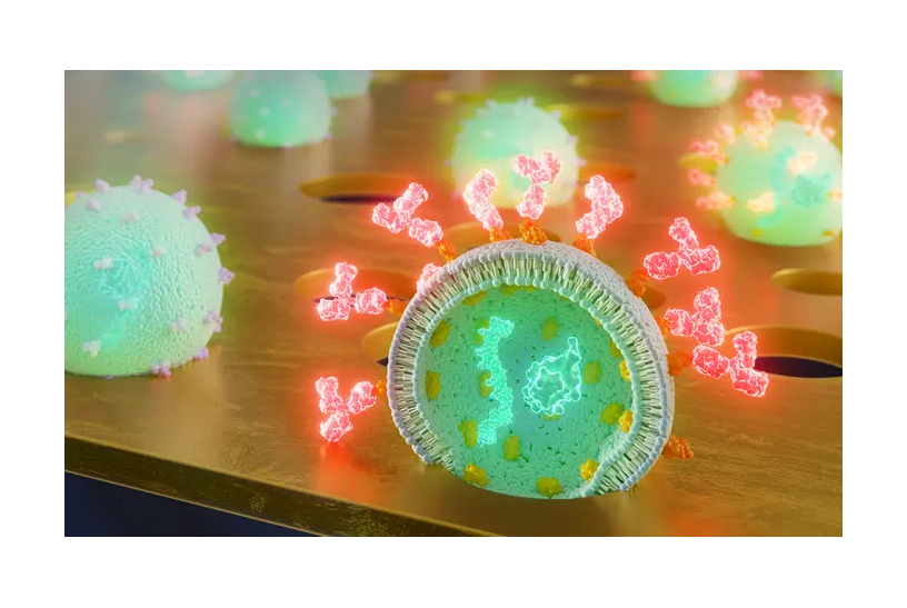
Featured image: A team of researchers at the University of Rochester has developed ultrathin membranes with pores sized perfectly to catch and display extracellular vesicles (EVs), tiny packets of cellular material that can provide important information about the status of the body. Photo: University of Rochester illustration / Erik Patak

