
A new identification reagent speeds diagnoses of chickenpox and shingles.
By Abbas Vafai, PhD, and Nicholas Vafai, PhD, MBA
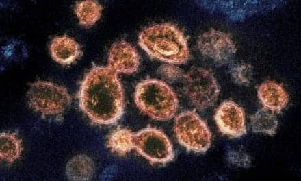
Varicella-zoster virus (VZV) is associated with two distinct diseases: childhood chickenpox (varicella) and shingles (herpes zoster). Shingles is caused by reactivation of VZV in elderly and immunosuppressed individuals. In the United States, there are an estimated 1.2 million cases of shingles each year.1
It is known that both VZV-specific cell-mediated immunity and neutralizing antibodies are crucial for the development and maintenance of immunity to VZV infection; waning cell-mediated immunity plays a role in the reactivation of VZV (zoster). Due to immune senescence, specific immune responses against VZV antigens may decline with age, and their capacity to prevent herpes zoster (shingles) will diminish. These factors result in an increased reactivation of zoster among aging and immunocompromised individuals.1–3
Following primary infection, the virus becomes latent in dorsal root ganglia and may reactivate years later to produce shingles in aging and immunocompromised patients, including transplant patients, children with cancer, patients taking steroids, or patients with AIDS. In both adults and children with AIDS, VZV may cause rare but clinically significant cutaneous, ophthalmic, and neural complications (Figure 1).3
Shingles Clinical Manifestations
The first symptom of herpes zoster (shingles) is usually pain, which may result from nerve damage caused when VZV reactivates from latently infected sensory neurons. Lesions erupt in the parts of the body where the pain is located (Figure 2). The pain, which can range from mild itching or tingling to sharply agonizing, is usually followed within 5 days by a vesicular rash within the infected dermatome (most commonly the thoracic, lumbar, or cervical regions) and clusters of clear vesicles, which soon develop into blisters.2,3
The etymology of the name ‘herpes zoster’ is explained by the clinical presentation of shingles. The name is derived from the Greek terms ‘herpein’ (to creep) and ‘zoster’ (girdle or zone). The eruption of herpes zoster is typically unilateral, does not cross the midline, and is limited to cutaneous innervation of a single sensory ganglia.3
Shingles lesions evolve slowly and appear as grouped vesicles rather than single lesions. They may be scattered in patches or may be so numerous that they form a continuous band. Generally, shingles lesions occur around the chest, abdomen, or eyes (ophthalmic zoster). Lesions rarely occur on the extremities. VZV reactivation may also occur without skin eruption, a condition known as ‘zoster sine herpete.’2
The course of shingles is usually dependent on the age and immunological status of the patient. Normally, new shingles lesions continue to appear for 2 to 3 days. Within 14 days, the lesions become pustular and crust. At this point, they no longer contain the virus.3
In immunocompromised patients, skin lesions may continue to appear for 2 weeks, and scabbing may not occur for as long as 1 month. Although the course of shingles is often benign in terms of mortality, severe and debilitating complications may occur, particularly among immunocompromised patients. Major complications associated with VZV reactivation include disseminated zoster, encephalitis, myelitis, pneumonitis, and postherpetic neuralgia. Immunocompromised individuals are more likely to experience complications from herpes zoster.2,3
Postherpetic neuralgia is one of the major complications of herpes zoster (shingles). In this condition, the patient continues to feel pain even after the shingles skin lesions have crusted. The pain is often severe in the areas where blisters occurred. The affected areas are also extremely sensitive to heat and cold. Postherpetic neuralgia is thought to result from nerve damage caused by the virus. Risk factors for the development of postherpetic neuralgia may include severity of pain, significant sensory impairment, and advancing age.2,3
Zoster encephalitis occurs most often in elderly adults with skin lesions in multiple dermatomes. The incidence of zoster encephalitis is low. A decline in cell-mediated immunity to VZV occurs with advancing age and may reflect selective immunodeficiency for the virus.2,4
Myelitis is a rare complication of varicella and zoster. Clinical features are characterized by stiffness and weakness of the legs. Most patients improve significantly over time; however, mild lower extremity stiffness and weakness may persist in a few patients.2
Disseminated herpes zoster often occurs in patients with depressed cell-mediated immunity caused by various underlying clinical conditions and their related treatments, including cancer, radiation therapy, cancer chemotherapy, organ transplants, and long-term use of systemic corticosteroids. Disseminated herpes zoster is characterized as a generalized eruption of skin vesicles occurring 7 to 14 days after the onset of shingles (herpes zoster).3
Herpes zoster pneumonia may develop in immunosuppressed patients, especially bone marrow transplant recipients and patients with chronic lung disease. Pneumonia usually occurs 2 to 7 days after the shingles rash appears.4
Diagnosis
VZV is diagnosed based upon its clinical presentation or through the use of laboratory techniques. Clinical diagnosis is often based on the presence of a vesicular eruption in a dermatomal (belt-like) pattern, characteristic prodrome, or patient history of exposure to the virus.2,3
Herpes zoster (shingles) skin lesions can occasionally mimic those resulting from infection with herpes simplex virus (HSV), and the two types can be difficult to differentiate clinically. Disseminated herpes simplex and disseminated zoster may also be indistinguishable clinically, leading to misdiagnosis if no confirmatory laboratory tests are performed. Cell culture cultivation of the virus from infected skin lesions or tissue samples has been the principal method for diagnosing VZV infection (Figure 3). Immunofluorescent antibody assays are commonly used for the detection of membrane viral antigens. Each of these techniques has been used for the diagnosis and differentiation of HSV and VZV infections.2,3
In the past, the Tzanck smear was often used to detect multinucleated giant cells or inclusion-containing cells. However, the Tzanck preparation has a low sensitivity compared with newer techniques (60%), and cannot distinguish between VZV and herpes simplex. Detection of the virus in cell culture generally takes longer with VZV than with herpes viruses.
Among the serologic tests used to differentiate between VZV and other viruses, enzyme-linked immunosorbent assays, indirect fluorescent antibody tests, fluorescent antibody membrane antigens, and latex agglutination are the most common. However, serologic tests may be of limited utility because diagnosis must usually be made within a short period. In addition, heterologous antibody responses to herpes simplex and VZV may occur in some patients because the two viruses share common antigens. Nevertheless, serologic tests can be used to determine immune status or confirm diagnoses already made.3
Molecular virology techniques, including polymerase chain reaction (PCR)-based tests, have also been used for the diagnosis of both HSV and VZV infection. PCR techniques can be used to detect VZV DNA from skin lesion exudate or from cerebrospinal fluid. However, molecular techniques are more costly than immunofluorescent antibody assays, and require expensive laboratory equipment and personnel training.3
Treatment of Shingles
The drug acyclovir is widely used for the treatment of herpes zoster. Acyclovir reduces healing time, the appearance of new vesicles, the duration of pain, and the duration of viral shedding.
Other drugs have also been shown to provide adequate treatment of herpes zoster, including aciclovir (Zovirax), famciclovir, penciclovir (Denavir), valaciclovir (Valtrex), and all variants of acyclovir. Oral famciclovir (500 mg or 750 mg three times daily for 7 days) is an effective and well-tolerated therapy for herpes zoster.3
VZV Identification Reagent
Viro Research, Snellville, Ga, has developed Zostergent, a rapid test to detect VZV in skin lesions and tissue samples of shingles patients (Figure 4). Zostergent features six unique and highly specific monoclonal antibodies against VZV early and late proteins. The single-step, single-solution fluorescent antibody test can be performed in 20 to 30 minutes to detect and confirm diagnoses of chickenpox (varicella) and shingles (zoster) in clinical specimens and inoculated cell cultures.1,5,6
The test is supplied as a 3-mL solution containing Evans blue counterstain in a self-contained, single-step, single-solution detection system. Users cover the sample to be tested with one drop of solution, incubate at 37° C for 30 minutes, wash in PBS and water, air dry, mount, and observe under a fluorescent microscope.
Zostergent is prepared with fluorescein isothiocyanate (FITC), conjugated and mixed with VZV-affinity purified murine IgGs directed against VZV glycoproteins gE, gB, gH, gI, and viral immediate early proteins encoded by VZV gene 62 (VZV IE62).7
In a retrospective study involving 54 frozen samples, Zostergent was found to have 100% specificity (n=15; 95% CI 78.2% to 100%) and 100% sensitivity (n=39; 95% CI 91.0% to 100%) when examined by cell culture confirmation (Table 1). Overall agreement was 100% (54/54).
In the study, Zostergent was used to examine 39 different VZV clinical isolates, along with 15 different clinical isolates: five each of herpes simplex virus type 1 (HSV-1), herpes simplex virus type 2 (HSV-2), and cytomegalovirus (CMV). All 39 different VZV isolates exhibited positive fluorescence staining, whereas negative results were obtained with all HSV-1, HSV-2 and CMV isolates. Human fibroblast cells were included in each test as negative control cell cultures. Staining of uninfected human fibroblast cells with Zostergent exhibited negative results.
Zostergent sensitivity and specificity were also found to be 100% when examined by testing vesicle smears. A total of 48 vesicle smear specimens were tested. Of that total, 28 specimens (10 frozen, 18 fresh) were tested from patients with varicella or zoster diagnosed by clinical presentation or by virus isolation. Relative sensitivity of Zostergent was 100% (28/28; 95% CI 87.7% to 100%).
In addition, 20 specimens (9 frozen, 11 fresh) from 10 patients with HSV-1 and 10 patients with HSV-2 infections were tested and found negative when stained with Zostergent (Table 2). Relative specificity was 100% (20/20; 95% CI 83.2% to 100%). Crossreactivity of Zostergent with other viruses was also found to be negative as determined by direct fluorescent antibody testing using virus-infected cells (Table 3).
Crossreactivity was determined by direct fluorescent antibody test using virus-infected cells and the Viro VZV identification reagent. The following viruses were tested and found to be negative by the Viro VZV identification reagent: cytomegalovirus (CMV), Epstein-Barr virus (EBV), herpes simplex virus type 1 (HSV-1), herpes simplex virus type 2 (HSV-2), human herpes virus 6 (HHV-6), influenza virus types A and B, measles virus, parainfluenza virus types 1, 2, and 3, and rubella virus. In addition, the following cell lines demonstrated no crossreactivity with the Viro VZV identification reagent: African green monkey kidney cells (VERO), human embryonic lung fibroblast cells (MRC-5 and WI-38), human foreskin fibroblast cells (HFF), and human lung carcinoma cells (A549).
Conclusion
Shingles (herpes zoster) is caused by reactivation of VZV in elderly and immunosuppressed individuals. There are an estimated 1.2 million cases of shingles each year in the United States.
The incidence of shingles may continue to increase as the US population ages. Therefore, there is a need for a rapid and sensitive diagnostic test for the detection of VZV in shingles patients.
Viro Research has developed Zostergent, a rapid test to detect varicella-zoster virus in the skin lesions and tissue samples of shingles patients. Zostergent offers six unique and highly specific monoclonal antibodies against VZV early and late proteins. The test is a single-step, single solution fluorescent antibody test that can be performed in 20 to 30 minutes, in order to detect and confirm chickenpox (varicella) and shingles (zoster) in clinical specimens and inoculated cell cultures. l
Abbas Vafai, PhD, is senior scientific adviser at Viro Research. While on the faculties of the University of Colorado and the University of Illinois, he discovered a glycoprotein (gE protein) found on the surface of the varicella-zoster virus that could be used as a general vaccine antigen, enabling vaccine developers to avoid using the actual virus. The resulting vaccine is now marketed by GlaxoSmithKline under the brandname Shingrix. Vafai later served as branch chief and associate director at the Centers for Disease Control and Prevention, before retiring in 2015.
Nicholas Vafai, PhD, MBA, is founder and chief executive officer of Viro Research. In 2012, he cofounded and coinvented the patented technology behind Lipid Nanotechnologies, a company that specializes in commercializing lipid microarray technology for personalized medicine and miniaturizing high-throughput drug screening. In 2019, he established Viro Research, a company that strives to provide high quality, innovative, safe, and effective in vitro diagnostic devices for the detection of human herpes virus antigens and antibodies.
REFERENCES
1. Vafai A. Varicella-zoster virus subunit vaccine. The Open Vaccine Journal. 2009;2:17–27; doi: 10.2174/1875035401002010017.
2. Gilden D, Vafai A. The neurologic complications of varicella-zoster infection. In: Vinken PJ, Bruyn GW, Klawans HK, eds., Handbook of Clinical Neurology. Amsterdam: Elsevier Biomedical Publishers, 1989.
3. Vafai A, Berger M. Zoster in patients infected with HIV: a review. Am J Med Sci. 2001;321(6):372–380; doi: 10.1097/00000441-200106000-00003.
4. Gilden DH, Murray RS, Wellish M, Kleinschmidt-DeMasters BK, Vafai A. Chronic progressive varicella-zoster virus encephalitis in an AIDS patient. Neurology. 1988;38(7):1150–1153; doi: 10.1212/wnl.38.7.1150.
5. Vafai A. Antibody-binding sites on truncated forms of varicella-zoster virus gpI(gE) glycoprotein. Vaccine. 1994;12(14):1265–1269; doi: 10.1016/s0264-410x(94)80030-4.
6. Berger M, Shankar V, Vafai A. Therapeutic applications of monoclonal antibodies. Am J Med Sci. 2002;324(1):14–30; doi: 10.1097/00000441-200207000-00004.
7. Zostergent VZV identification reagent [instructions for use, online]. Snellville, Ga: Viro Research, 2019. Available at: https://viroresearch.com/wp-content/uploads/2019/08/viro-vzv-id-reagent-product-insert-030819.pdf. Accessed June 15, 2020.
Featured image: Shingles lesions caused by reactivation of varicella-zoster virus from sacral sensory ganglia.


