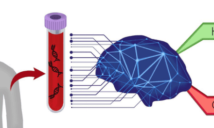

A study found that a blood test measuring cancer-associated proteins detected signs of early-stage ovarian cancer in samples collected up to a year before diagnosis.
By Alyx Arnett
Detecting ovarian cancer before symptoms develop has long been a challenge, particularly because existing blood-based tests can miss cancers in their earliest stages. Now, a study published in Gynecologic Oncology suggests that measuring proteins on particles released by living cells could offer another way to identify the disease before diagnosis.
Developed by Mercy BioAnalytics, the blood test measures cancer-associated proteins on extracellular vesicles and particles (EVPs). Researchers evaluated the test using blood samples collected before women were diagnosed with ovarian cancer, comparing its performance with CA-125, a commonly used ovarian cancer blood marker.
“We wanted to know whether the test detects signal associated with high-grade serous ovarian cancer in blood collected before anyone knew the woman had cancer,” says Toumy Guettouche, PhD, chief scientific officer at Mercy BioAnalytics and a co-author of the study.
The company’s Evident ovarian cancer screening test uses its Mercy Halo platform and has received FDA Breakthrough Device Designation. In this Research Q&A, Guettouche discusses the study’s findings, how the test works, and what additional evidence is needed to establish its clinical utility.
[Editor’s note: Read the study, “Extracellular vesicle and particle-based blood test for ovarian cancer screening of average risk population: a promising nested case control study using preclinical samples,” in Gynecologic Oncology]
What prompted you to explore EVPs as biomarkers for early ovarian cancer detection?
I came to Mercy BioAnalytics skeptical of extracellular vesicles (EVs) because the field was still evolving and assay reproducibility had been a challenge. Mercy works with a broader category: extracellular vesicles and particles (EVPs). Every EV is an EVP, but not every EVP is an EV. Methods that collect vesicles from blood also collect membrane-free particles, and both can carry cancer signals. I quickly recognized that EVPs could address a fundamental limitation of liquid biopsy that I had experienced firsthand.
Earlier in my career, I worked on ctDNA-based diagnostics and saw how challenging it was to detect early-stage cancers when only tiny amounts of tumor-derived DNA were present in circulation. EVPs offered a different opportunity. Even small tumors can release large numbers of these particles, each carrying molecular information from its parent cell. I think of them as miniature biopsies that allow us to interrogate multiple tumor-associated biomarkers simultaneously, potentially providing a much richer signal even at the earliest stages of disease.
Can you walk us through the study and what you were hoping to learn from evaluating preclinical blood samples?
The preclinical samples—collected before the women were diagnosed with cancer—came from UKCTOCS, the United Kingdom Collaborative Trial of Ovarian Cancer Screening, which randomized around 200,000 women. We ran a blinded case-control study nested within it, drawing on the ultrasound and no-screening arms. Forty-one women later diagnosed with high-grade serous cancer were matched by age to 1,176 controls, and the primary analysis used samples collected up to 12 months before diagnosis.
Preclinical blood is much closer to what a screening test sees in practice than blood drawn at diagnosis, when the tumor is already known. The study is possible because EVPs remain stable in samples banked for decades once they are properly processed. Archived cohorts answer the detection question in months rather than years. They cannot answer whether screening changes outcomes, which is why the prospective work or modeling studies still have to be done.
What were the most important findings?
For Stage I/II high-grade serous ovarian cancer, in samples collected up to 12 months before diagnosis, the test detected eight of nine cases, corresponding to 89% sensitivity, compared with five of nine for CA-125. Specificity was approximately 98% across 1,176 controls. The assay is designed to improve specificity by requiring multiple cancer-associated proteins to be present on the same particle, helping distinguish cancer-associated signals from background signals originating from healthy tissue.
For a screening test, the balance between sensitivity and specificity is critical, and the study demonstrated strong performance on both measures. Importantly, the study demonstrated the potential to detect cancer-associated signals well before clinical diagnosis, including one case detected approximately three years earlier in the longitudinal analysis.
Was there anything in the results that surprised you?
The surprise was how much a 20-year-old sample can still tell you. EVPs tend to survive long-term storage, and the signal distributions we see in freshly collected blood look like those in samples banked two decades ago. That means we can go back to screening trials where women gave blood every year and ask when the test first turns positive relative to clinical diagnosis.
It also lets us separate something the field usually runs together. The first screening round flatters every test because it picks up cancers that have been accumulating undetected for years. Later rounds are harder because the cancers appearing then are newer and smaller. Most published screening sensitivities are first-round numbers, so the benchmarks the field works against are systematically optimistic. Archived longitudinal cohorts let us measure that gap now, instead of waiting years for prospective follow-up. That has changed how I read any screening performance claim, including our own.
How does measuring tumor-associated biomarkers on EVPs differ from other blood-based approaches?
Different blood-based tests read different biological signals, and the difference matters most in early disease. ctDNA reaches the blood largely when tumor cells die, so the amount available scales with how much tumor is dying. In a small tumor, that can mean very few copies in a tube, a limit no assay sensitivity can overcome. EVPs are released continuously by living cells, including cells in small tumors, so the signal does not depend on cell death. They are also abundant and stable in storage. The challenge is proportion rather than shedding rate. A tumor cell releases more EVPs than a healthy cell does, but early in disease, healthy cells outnumber tumor cells by orders of magnitude, so they dominate the circulating pool.
Many cancer-associated proteins are also produced by healthy or benign tissues, which is why single-analyte blood tests like CA-125 struggle with specificity. The same holds true on vesicles: most of the tumor-associated proteins we target also appear, at lower levels, on vesicles from healthy or benign cells. A healthy or benign vesicle may carry one of these proteins but rarely carries several at once; a tumor-derived vesicle is far more likely to. Our assay therefore requires multiple cancer-associated proteins on the same vesicle, a design intended to separate tumor signal from healthy or benign background and improve signal-to-noise at early stages.
For clinical laboratorians, what is important to understand about how the assay identifies and measures the ovarian cancer-associated signal in a blood sample?
The concept to understand is colocalization. Mercy Halo captures EVPs from the blood sample on antibody-coated beads, then asks whether several cancer-associated surface proteins sit on that same individual EVP. Approaches that measure each protein separately cannot ask that question. The method is what lets it run on standard equipment. The detection antibodies each carry a short oligonucleotide. Those oligonucleotides can ligate only when both antibodies are bound to the same EVP, and the ligated product is what qPCR amplifies. An EVP carrying only some of the required markers is designed not to generate a template, and therefore no signal.
For a laboratory, this means specificity is built into the chemistry rather than recovered afterward. Background from healthy or benign cell EVPs is suppressed at the point of detection, so the algorithm and cutoff work on a low-background signal rather than compensating for a noisy one. Background is not zero: A small fraction of healthy or benign EVPs carry all three markers, so the cutoff separates a low baseline from tumor signal. The readout is quantitative PCR on standard laboratory instruments, with no sequencing and a semi-automated workflow, and the assay has been run on both serum and plasma.
What challenges need to be addressed before an EVP-based assay like this could be used for routine ovarian cancer screening?
Evident, our ovarian cancer screening test, which is not FDA cleared or approved, runs on this platform in our CLIA laboratory. The question is what routine EVP-based screening requires. First, reagents. These assays depend on antibodies of widely varying quality, and coverage of the surface proteome remains incomplete.
Second, sample handling. Evident runs on serum, processed on site and shipped at room temperature the same day. EVPs are typically stable once processed, even in samples banked for decades. The window before processing must be controlled, and holding that standard across many sites is the challenge at scale.
Third, the care pathway. A positive result can precede anything visible on imaging, typically transvaginal ultrasound. Clinicians and laboratories need an agreed plan for a positive test with a normal scan, such as repeat testing at a shorter interval rather than surgery.
Fourth, evidence from the intended-use population, showing how sensitivity and positive predictive value hold up across annual rounds. Mercy has two UKCTOCS studies using prospectively collected samples from this population: one published (Manning et al, Gynecol Oncol 2025) and one longitudinal study complete, with the manuscript in preparation. A prospective safety study is soon to start.
What are the next steps for the research, and what additional evidence will be needed to establish the clinical utility of this approach?
Validity and utility are different bars. The studies above establish how well the test detects cancer. Utility requires showing that using the test improves outcomes for women, weighed against the harms of false positives and the surgery they can trigger.
Our Evident ovarian cancer screening test is designed to screen postmenopausal women at average risk. FDA has approved our Investigational Device Exemption, allowing us to use Evident in SOARA, a prospective pivotal study in this population. The study will evaluate test- and screening-related adverse events over 12 months, determine specificity, and assess downstream diagnostic burden, including the screen-to-surgery ratio. SOARA is scheduled to begin once we receive Institutional Review Board approval.
We will continue to use archived samples from large screening trials to measure performance in incident screening rounds, where cancers arise after a prior negative screen and are typically smaller at detection. We also plan to assess Evident prospectively in symptomatic women undergoing evaluation for possible ovarian cancer through a clinical utility study planned for next year.
The need there is distinct. When a woman presents with an adnexal mass, the central question is whether it is malignant or benign. CA-125 and HE4 are often elevated in benign masses, so they separate the two imperfectly. Beyond ovarian cancer, we have a lung program in development, and our longer-term goal is multi-cancer detection at stages I and II.
ID 399475437 © Sampon Stu | Dreamstime.com
Do you want your research featured in a future CLP Research Q&A? Email [email protected].