
Whether in enabling precision medicine, innovating for quality assurance, or monitoring the effectiveness of therapy via companion diagnostics, ePathology tools bring pathologists closer to their patients

“Skate to where the puck is going, not where it’s been,”
Wayne Gretzky
1999 Inductee National Hockey League Hall of Fame
National Hockey League Hall of Fame player Wayne Gretzky always seemed to be in the right place at the right time. He was known for playing smart, anticipating what was needed, and putting himself in a position to provide it.
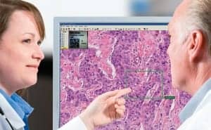
Efficient and effective approaches that sharpen the profile of a forward-looking laboratory rely upon ePathology tools that can enable pathologists and laboratory staff to anticipate and provide what is needed for optimal patient care. In a patient care environment served by a digital (or ePathology) laboratory, sophisticated support tools enable optimal patient care planning, and day-to-day decisions are facilitated by rapid electronic results.
I have visited many laboratories in the last decade. Invariably, I learn something. Individuals approach a challenge and adapt to change in a variety of instructive ways. And it is interesting to see how people see themselves on the arc of change.
For example, some pathologists have told me that they have been using digital pathology for 20 years. And they would be correct, if science were static and ePathology meant a digital photo taken with a camera hooked up to a microscope. But in today’s environment, that’s not the half of it.
Modern laboratories can be equipped to offer far more than the ability to capture images with a camera attached to a microscope. Today, ePathology can enable decision support when a pathologist seeks a consult to confirm his or her thinking. It is being used in training programs, exposing residents to an amazing library of images. It is employed in research, to record incremental progress and rapidly share new knowledge. Licensure examinations for newly-minted pathologists require ePathology tools as well.

JARED N. SCHWARTZ, MD, PhD chief medical officer, Aperio Inc, Vista, Calif, and consulting professor of pathology, Stanford University Medical Center, Palo Alto, Calif.
ePathology will not replace the microscope because it is the ideal tool in certain situations. But as technology evolves, those who use that technology evolve in tandem. Growing pains are aptly named; transition can be challenging; but at the end of the day, precision analytics and eSlides will allow us to go beyond the microscope to greater patient benefits.
Today, the use of ePathology will mean that companion diagnostics will sometimes dramatically impact patient care decisions, enhancing the likelihood of recovery. ePathology will enable us to monitor a patient’s response to certain treatments with a speed and certainty that was inconceivable a generation ago. The potential of ePathology continues to evolve; its limits are not yet apparent.
ePATHOLOGY AND THE EHR
One key goal in laboratory medicine is to integrate all patient records, an objective that requires all images to be in an electronic format. Integrating laboratory results in the EHR will enable the benefits of ePathology (improved transparency, consistency, and efficiency) to come to the foreground. Given that at least 70% of patient care decisions are tied to laboratory tests, including these images in the EHR makes their importance clear.
In evaluating a shift to whole slide imaging, consider the purposes that you might find for that image. Ask yourself if there is a problem that can’t be solved without using this technology—archiving/retrieval, sending consults, etc. Take a clear-eyed look at your challenges—today and in the future. If there are tasks that you cannot now do in-house, or inconveniences that are increasingly frustrating, what available tool could best solve your problem?
SHOW ‘EM, THEN TELL ‘EM
ePathology can increase access to pathologists and subspecialists.
Some people are uncomfortable with new technologies; they want to see them work. In this situation, collect a few case studies. Collect evidence of the need to upgrade and present the case for ePathology as a much-needed solution.
Here’s a true story. A pre-eminent pathologist adamantly refused to bring telepathology tools into his laboratory. Then, one winter weekend when he was on call, he had to drive through a blizzard to read a couple of slides for his resident. He came around after that … and never looked back. It’s like the couple who wanted nothing to do with Skype® … until their grandchildren moved across the country.
POTENTIAL FOR ePATHOLOGY TOOLS
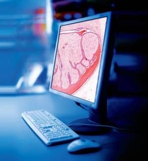
ePathology is about ease of consultation, ready access to companion diagnostics, archival of images, instantaneous retrieval of a complete EHR record, quantitative image analysis tools, and education. Consider the powerful potential of universal access to libraries of digitized slides provided by the best pathologists in the world. Think of the uses for a repository of these images. What would it mean to have thousands of slides representing unusual cases that can be searched and sorted in minutes? The value to education and research will be exponential.
Imagine that your physician referred your case to me for examination for a lesion of your lung. Then imagine that you had a skin biopsy 10 years before, which I realized upon reviewing your EHR. I would want to review the old pathology because it might reveal something that might explain a mass that might have been missed; perhaps a melanoma. Knowing that would affect treatment planning. Once upon a time, your skin biopsy would have been put into a file and stored off-site. We would need to request retrieval.
I once asked pathologists at a couple of medical centers in New York City about time required to retrieve tissue. “Two to three weeks if you’re lucky,” they said. “They’re stored in New Jersey.”
Most medical centers don’t store tissue samples on-site for more than 2 years because they take up a lot of space; it’s expensive. And you have to wonder, with all that material management, how often is a slide misfiled?
Now think about that same situation today, if ePathology were part of standard pathology practice. At the first visit, I would scan your tissue, interfaced the image with your EHR in the Laboratory Information System (LIS), and only then send the slide to storage. When you returned a few years later with a new problem, instead of making a request to “find the slide” and delaying your report on the consultation, scheduling a 3-week follow-up, I would have the ability to retrieve the first biopsy’s image and compare it then and there with the current specimen. The report would go out the same day, and care planning could begin.
Archival and retrieval of scanned biopsy images is already done in some US medical centers. The pathologist can review the old and new slides side by side on-screen. ePathology saves time, money, and perhaps most important, patient worry.
ePATHOLOGY IN THE LABORATORY
ePathology includes far more than anatomic pathology services. Laboratory medicine provides many potential opportunities to use the tools of ePathology—its chemistry, its hematology, its microbiology, its genomic. Those who work in the lab are already using many of these new technologies. Some of the most exciting potential uses for ePathology relate to genomics. Without electronic images and image analysis making precision medicine possible, the promise of genomics would be sadly limited.
A pathologist, a microscope, and a slide walk into a bar …
… and they all order ePathology. The pathologist wants it so he can obtain consults in minutes, store information in a more retrievable form, and integrate AP findings in the medical record. The microscope wants it because it knows that while it will always have a place at the table, yesterday’s table is today’s tablet. And the slide wants it because it was there at the beginning and wants to follow the technology wherever it takes it. The pathologist, microscope, and slide can agree: When time, flexibility, and retrievability are critical, the medium is key to the message. They all drink to success.
Just like computers and smartphones, ePathology will continue to get better and better. As physicians, medical technologists, cytotechnologists, and histotechnologists, we are focused on how these tools will enable us to solve problems that compromise our efficiency and limit our effectiveness in improving patient outcomes.
Of all the benefits that will accrue as ePathology moves forward, improved access to care for those in remote areas will be one of the most significant. ePathology can make a dramatic difference in quality of life for many thousands of people in rural and remote locations in the United States, as well as around the globe, in places where remote populations are without easy access to pathology services.
ePATHOLOGY AT THE RIGHT PLACE, AT THE RIGHT TIME
I opened this story with a quote from Wayne Gretzky, who played 20 seasons in the National Hockey League. Gretzky wasn’t especially big or aggressive, but he understood the value of anticipation.
Ten years from now, someone who had worked in the laboratory in 2012 might not recognize what pathology will have come to be. Pathologists and other laboratory staff are anxious to further integrate the information that we get from various parts of the lab, be it anatomic, molecular, or chemistry labs within a hospital system, and a truly comprehensive patient medical record. We continue to jump the hurdles of the day and look for the next ones, knowing that our work is truly central to quality health care and patient safety. Laboratory professionals understand better than most the fundamental importance of quality measures. And we are ideally positioned to know—and show—where the puck is going.
The statements, views, and opinions expressed in this article are those of Jared N. Schwartz, MD, PhD. Dr Schwartz has more than 30 years of experience in the field of pathology. He served as president of the College of American Pathologists, and director of pathology and laboratory medicine at Presbyterian Healthcare, Charlotte, NC. Board certified in anatomic and clinical pathology with subspecialty boards in medical microbiology and cytopathology, he is a graduate of Duke University Medical School, where he completed his residency and fellowship training, and served as chief resident. Schwartz was appointed to the Clinical Laboratory Improvement Advisory Committee by HHS, and was a co-chair and author of the ASCO/CAP Guidelines on HER2, which was published in the January 2007 editions of the Journal of Clinical Oncology and Archives of Pathology and Laboratory Medicine. For more information, contact Editor Judy O’Rourke, .
ANATOMIC AND DIGITAL PATHOLOGY RESOURCES:
- Aperio: www.aperio.com
- Biocare Medical LLC: www.biocare.net
- Cerner Corp: www.cerner.com/laboratory
- ChemImage Corp: www.chemimage.com
- Cortex Medical Management Systems: www.cortexmed.com
- Definiens: www.definiens.com
- LigoLab Information Systems: www.ligolab.com
- 4medica: www.4medica.com
- Hamamatsu: sales.hamamatsu.com
- Leica Microsystems: www.leica-microsystems.com/products/digital-pathology
- Milestone: www.milestonemed.com
- NovoPath: www.novopath.com
- Olympus America: www.olympusamerica.com
- Omnyx: www.omnyx.com
- Orchard Software Corp: www.orchardsoft.com
- PathCentral Inc: www.pathcentral.net
- Sunquest: www.sunquestinfo.com
- Thermo Scientific Anatomical Pathology: www.thermoscientific.com/pathology
- Ventana Medical Systems Inc: www.ventanamed.com

