Device detects inflammatory signals within minutes and collects specialized immune cells within hours, offering alternative to blood draws and biopsies.
Researchers at The Jackson Laboratory (JAX), in collaboration with the Massachusetts Institute of Technology (MIT), have developed a bandage-like microneedle patch that can sample the body’s immune responses painlessly from the skin without requiring blood draws or surgical biopsies.
The device detects inflammatory signals within minutes and collects specialized immune cells within hours. The study appears in Nature Biomedical Engineering.
“Traditionally, studying some of the most important immune cells in the body requires a skin biopsy or blood draws. Because many of these cells live and respond in tissues like the skin, accessing them has meant invasive procedures,” says Sasan Jalili, PhD, a biomedical engineer and immunologist at JAX, in a release. “We’ve shown we can capture them painlessly and noninvasively instead.”
Leveraging Resident Memory T Cells
The patch works by harnessing resident memory T cells, immune sentinels that live in skin and other barrier tissues and rapidly respond to previously encountered foreign threats, or antigens. When these cells recognize a familiar antigen, they release signals to attract additional immune cells from the bloodstream, including highly specialized T cells that recognize that same threat.
By triggering this natural process, which concentrates key immune cells in the skin, the researchers assessed immune responses. The sampled material revealed the number and state of T cells and other signaling molecules, offering a readout of the immune system’s strength and responsiveness to specific diseases and conditions.
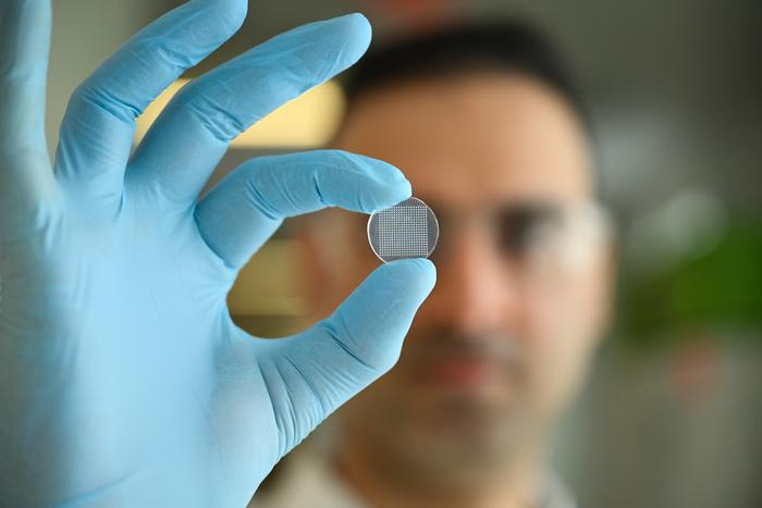
The patch contains hundreds of microscopic needles made of a Food and Drug Administration (FDA)-approved polymer. A seaweed-derived hydrogel also deemed safe by the FDA coats the needles and absorbs immune cells and molecules from skin interstitial fluid. The microneedles reach only the upper skin layers, causing minimal irritation and no damage to nerves or blood vessels.
Clinical Testing Results
In mouse vaccination models, the patch dramatically boosted the recovery of antigen-specific T cells, recruiting many of these cells from the bloodstream rather than skin. In a human test at University of Massachusetts Chan Medical School, the patch also collected immune cells and signaling proteins, including resident memory T cells.
“This study marks the first demonstration of live human immune cell sampling using a microneedle patch,” says Jalili, who is also a joint faculty member at University of Connecticut School of Medicine, in a release. “This opens the door to a new way of monitoring immune responses that’s practical, painless, and clinically feasible.”
Clinical Applications
The patch is already helping researchers and clinicians study immune responses in aging and skin autoimmunity, including vitiligo and psoriasis. The technology could make it easier to track how people respond to vaccines, infections, and cancer therapies by complementing traditional blood tests and biopsies.
The patch may be especially useful for skin conditions, since immune cells that drive conditions such as allergic dermatitis, psoriasis, and vitiligo already live in the tissue. Jalili is using it to study how age-related skin changes contribute to chronic inflammation and frailty in older adults.
“Not only did we run extensive preclinical experiments, we were able to carry out an initial test in humans,” says study co-author Darrell Irvine, PhD, an immunologist and bioengineer at Scripps Research, who began the work at MIT, in a release. “That’s exciting because it almost never happens with brand-new technologies.”
Looking ahead, the patch could eventually support at-home monitoring, allowing patients with skin conditions to track unpredictable flare-ups. The technology could also be adapted for oral or nasal cavities, opening the door to monitoring mucosal immune responses.
“People wouldn’t need hours of sampling. Even 15 to 30 minutes can be enough to detect inflammatory signals and get a sense of what’s happening in the tissue,” says Jalili in a release.
Photo caption: Sasan Jalili holds the microneedle skin patch, which is about the size of a quarter.
Photo credit: The Jackson Laboratory


