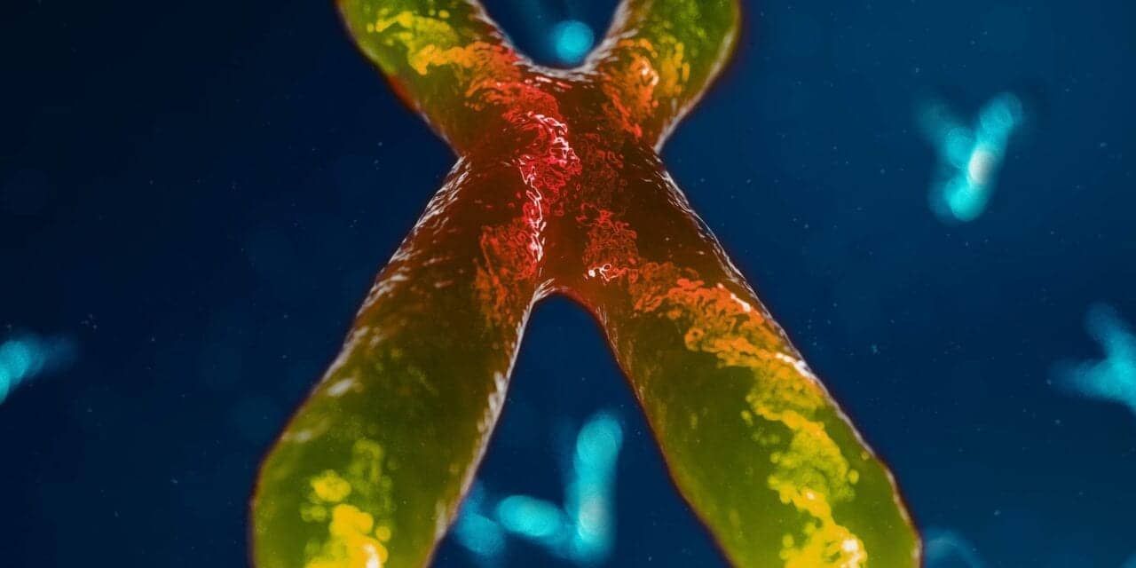
Fluorescence in situ hybridization (FISH) has been the tried and tested way to visualize and map genetic material in a cell, but advances in technology are making this traditional testing method more efficient, cost effective, and robust.
By Brian Kirk
Cytogenetic testing is often associated with tried and tested technologies that are, perhaps, a little out of date. But modern medicine still relies heavily on these methods for the diagnosis of a wide range of conditions. This is because, despite numerous technological advances in genetics and next generation sequencing (NGS) over the past 20 years, the cost and complexity of whole genome sequencing mean that cytogenetic testing is still far from being the gold standard for the detection of genetic abnormalities.
Cytogenetic analyses typically involve the microscopic examination of patient samples to identify chromosomal abnormalities and are commonplace for the detection of genetic disorders and hematological diseases, as well as for diagnosis and monitoring in oncology. Unlike karyotyping, which relies on the availability of a fresh sample of proliferating cells, fluorescence in situ hybridization (FISH) works with fixed cells or tissue samples, bypassing the need for a cell culture step. Instead, it uses fluorescent probes targeting specific DNA sequences to detect known genetic alterations via standard laboratory methodologies.
Old FISH, New FISH
FISH provides an effective way to visualize and map the genetic material in a cell. Scientists can detect genetic abnormalities – deletions, duplications, or translocations – with fluorescent probes, and can even perform so-called chromosome “painting,” using multiple fluorophores to detect chromosome rearrangements. This technology is the chosen tool for evaluating many biomarkers due to its simplicity and reliability, and has been a key platform for monitoring disease progression in cancer patients. It is also used extensively in research labs to help further our understanding of disease biology.
While FISH has traditionally consisted of a more manual, hands-on approach, its high specificity and accuracy, together with the many protocol options and the ability to use a broad range of sample types – from blood to solid tumors – still make FISH the gold standard technique for the detection of genetic abnormalities.
There have certainly been attempts to replace FISH with technologies such as NGS in a clinical setting, but these efforts have not been as successful as initially expected. This lack of efficacy is due largely to a combination of the relatively high costs of NGS, the reluctance of busy clinical labs to completely transform their workflows to incorporate novel technologies, and the need to recruit expert bioinformaticians who can analyze the large quantities of data generated using NGS in a meaningful way.
For these labs, it makes more sense to update FISH technologies than to introduce new techniques. They can rely on expertise and know how already present in the lab, as well as the existing robust reagent supply chain, while avoiding the need for dramatic changes to workflows. For many labs, the decision to use FISH is clear cut. Why develop a new technology from scratch, when there is already an established and validated one as a good foundation?
Changing the paradigm
Bringing clinical cytogenetics into the 21st century requires a change in the way labs approach FISH. Instead of a one patient, one sample – and a reliance on a manual – workflow, labs will need to adopt efficient, high throughput approaches to make the process faster and more cost effective.
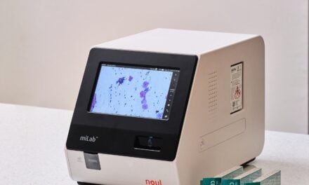
For example, Irvine, Calif.-headquartered BioDot, Inc., has taken on the task of revamping FISH to offer a more efficient, multiplexed platform. The company has used its proprietary, non-contact, and quantitative fluid dispensing technology – essentially an inkjet printing system for biomolecules – to miniaturize and automate slide preparation. Capable of dispensing nanoliters of fluid onto glass slides with increased precision, CellWriter technology addresses many of the inefficiencies of classical FISH by enabling reliable testing of much smaller sample sizes. It enables reproducible assays to be performed using around 200 cells per sample, with eight individual samples per slide. This reduces the volume of expensive fluorescent probes required from 10 microliters per test to just 0.5 microliters per sample, dramatically cutting the cost per patient. Other benefits of this automated approach are that slides are prepared faster, without the need for constant user oversight, while generating fewer pieces of glass to process and analyze.
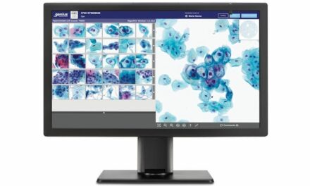
Standardization is another key factor in improving workflow efficiency, and this improved FISH methodology can be used to process various matrices, diluting or concentrating samples automatically prior to printing them onto the slides. The increased reproducibility offered by this approach also enables reliable automation of analyses, using an automated fluorescence microscope to further reduce turnaround times. Similarly, a built-in barcode reader means that each sample is carefully tracked from collection to result, making sample management a lot easier and far more efficient. Crucially, this approach is vendor-independent, and has been validated for most of the common FISH applications, using a wide range of different probe sets.
The Future of FISH Is Now
Standardization, automation, and miniaturization are bywords for increasing efficiency and throughput in a laboratory environment, and the simple, liquid-based chemistry and immunoassay workflows in clinical diagnostics labs have already undergone this transformation.
Cytogenetics has, until now, lagged behind other disciplines, due to a lack of accurate, reliable and reproducible technologies to automate processing. New technology addresses these challenges to bring FISH workflows into the 21st century, creating a simple, cost-effective, and automated process that will keep FISH as the gold standard for cytogenetic testing and benefit patients for years to come. Welcome to Cytogenetics 2.0.
About the Author
Brian Kirk is vice president for Business Development at BioDot, Inc., and has over 15 years of experience designing, developing, and marketing high-throughput manufacturing systems for many of the world’s largest diagnostic companies. He led the development and commercialization effort to disrupt the FISH and cytogenetics markets with the patented FISHArray technology. He now works with groups in life science (high-throughput screening, genomics, proteomics, etc.), leveraging BioDot’s expertise in nanoliter and picoliter dispensing to build next generation tools in these areas.
