
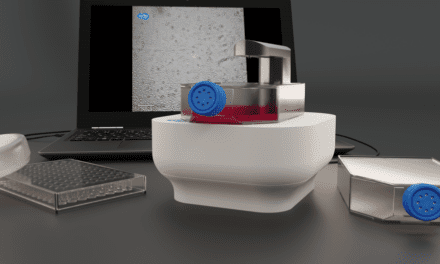
Yokogawa Electric Corp. unveiled a new high-content analysis system for capturing high-definition 3D microscopic images of live cell cultures. Expanding the company’s CellVoyager family of products, the CQ3000 will be launched commercially later in 2024.
The CQ3000 has been designed to capture 3D microscopic images of live cell cultures in high definition at high speed. When used together with Yokogawa’s CellPathfinder image analysis software, the CQ3000 can quantify and analyze intracellular organelles to assess cellular reactions and the effects of drug compounds, the company says. It enables highly efficient evaluation of cells in a wide range of applications, from basic research to drug discovery screening.
“The CQ3000 is an enhancement to the CellVoyager series lineup that will accelerate the development of new drugs and streamline basic research in cutting-edge biology and medicine,” says Hiroshi Nakao, Vice President and head of the Life Business Headquarters at Yokogawa Electric. “Through the provision of products and solutions that contribute to drug discovery research and the advancement of individualized medicine and regenerative medicine, Yokogawa’s aims to ensure well-being and quality of life for all.”
About CQ3000
The CQ3000 enables the stable and precise observation of live cell cultures over long time periods thanks to Yokogawa’s high-precision temperature control and dual spinning-disk with microlens that captures optical slice images (confocal images) with higher speed and better sensitivity, which minimizes damage to cells compared to other confocal systems on the market, the company says.
The CQ3000 can capture images of microplates at high speed thanks to enhancements to its stage control and auto-focus functions. When used in combination with optional dual camera and wide-field image capture feature, which uniformly captures the entire field of view, image capture time is reduced. This product enables the high-speed selection of promising chemical compounds from vast numbers of new drug candidates, according to Yokogawa.
By using Yokogawa’s proprietary water immersion lens mechanism, in addition to detailed observation at high magnification levels, it is also possible to capture bright images at low magnification levels. The use of an optional uniformizer achieves even illumination across the entire field of view, for the capture of vivid images.
Further reading: Molecular Imaging & Histology Combine for Better Cancer Diagnosis
Photo: Yokogawa Electric


