
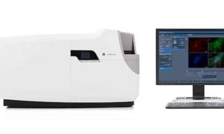
Leica Microsystems, Mannheim, Germany, has launched Stellaris, its new flagship confocal microscopy platform, designed to significantly improve how scientists are able to capture three-dimensional images of living cells and tissues.

Root-hypocotyl junction of Arabidopsis thaliana. Simultaneous spectral (gray) and TauSense mode confocal imaging with TauContrast (color) reveals contrast by lifetime. Image by Melanie Krebs, PhD, courtesy University of Heidelberg.
Due to its optimized imaging performance, the new confocal platform gives researchers the power to see more and, as a result, collect more accurate and reliable data to prove hypotheses with precision. The combination of the unique new power HyD detectors, white light laser, and sophisticated software delivers brighter signals, resulting in images with more contrast and astounding detail. Such data help researchers to observe the inner workings of cellular processes—crucial for research in fields like cancer and neuroscience.

Markus Lusser, Leica Microsystems.
“Stellaris offers additional information that is simply not available in conventional confocal systems,” says Markus, Lusser, president of Leica Microsystems. “You get immediate access to functional information without having to learn complex, advanced techniques. This development will open up confocal microscopy to a much wider group of scientists and researchers, drive up productivity, and enable new breakthroughs to be achieved in laboratories around the world.” Stellaris replaces Leica Microsystems’ SP8 platform with two main systems: the Stellaris 5 and the advanced Stellaris 8. For more information, visit Leica Microsystems.
