
ZEISS unveiled two new software functionalities to help medical professionals achieve better results in confocal microscopy, the ZEISS LSM Plus and ZEISS Airyscan Joint Deconvolution.
ZEISS Laser scanning microscopes (LSMs) provide a wide range of detection modes with highest sensitivity and spectral flexibility for multi-fluorescent experiments. ZEISS LSM Plus and ZEISS Airyscan Joint Deconvolution are designed to improve the data quality in confocal imaging. This enhancement is independent of the chosen detector, imaging mode, and emission range.
The existing ZEISS LSM 900, including ZEISS Celldiscoverer 7, and ZEISS LSM 980 microscope systems can be upgraded accordingly with the new software functionalities.
The linear Wiener filter deconvolution of the new ZEISS LSM Plus option is based on the super-resolution processing of ZEISS Airyscan. It can be applied to data of any confocal imaging mode with next to no interaction, ensuring reliable quantitative results. Users benefit from an enhanced signal-to-noise ratio at high acquisition speed and low laser power—particularly useful for live cell imaging of delicate samples. For samples that are not too light sensitive and therefore allow the pinhole of the LSM to be closed, more spatial information and even greater resolution enhancement can be obtained.
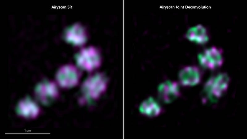
Meanwhile, the ZEISS Airyscan 2 is an area detector with 32 circularly arranged detection elements. Each of these acts as a small pinhole with a slightly different view on the sample, providing additional spatial information. With the introduction of ZEISS Airyscan Joint Deconvolution, this additional spatial information can now be leveraged to reduce the distance that can be resolved between two points down to 90 nm. Super-resolution experiments benefit from an improved separation of single or multiple labels.
Featured image: Cos-7 cells imaged with ZEISS LSM Plus. The fluorescent signals were separated by Linear Unmixing, facilitating clear separation between the spectrally overlapping dyes. Photo: ZEISS
