The diagnostic gold standard in most surgeries is the extraction of biopsies and their histopathological examination to confirm the tumor and the tumor borders. The histopathological examination is carried out by means of rapid sections of non-contrasted tissue sections during the surgery and also on tissue sections of fixed material. This procedure is time-consuming; investigator-dependent; and dependent on the size, number, and quality of the removed tissue samples. Thus, in tumor surgery there is a great need for new technologies for in vivo tissue diagnostics so that surgeons can remove it as completely as possible, because a reliable detection of tumor margins is the key to effective tumor treatment.
In this context multimodal nonlinear imaging combining the three different nonlinear imaging techniques—namely coherent anti-Stokes Raman scattering (CARS), two-photon excited autofluorescence (TPEF), and second harmonic generation (SHG)—offers the potential to reliably assess tissue and the success of surgery or endoscopy. However, the in vivo tissue diagnostics application of these methods during endoscopic or surgical procedures is challenging requiring (i) robust ultrafast lasers, (ii) low loss laser delivery and signal collection fibers maintaining the pulse shape, (iii) compact, fast and precise scanners as well as (iv) high-performance endomicroscopic objectives.
In a new paper published in Light Science & Application, a team of scientists from Jena, Germany, led by Juergen Popp, PhD, of the Leibniz Institute of Photonic Technology and Friedrich Schiller University Jena and Bernhard Messerschmidt, PhD, of the company Grintech introduces a novel all-fiber based endoscopic set-up for multimodal non-linear endoscopy allowing to record in vivo tissue diagnostics images displaying both morphological and biochemical information.
The scientists have developed a CARS/SHG/TPEF endoscopy platform, in which they have custom designed all abovementioned key components for best performance, i.e., the portable fiber laser, a new type of solid fiber for guiding the excitation laser wavelengths in two separate cores and collecting the signal in an outer collection cladding, a resonant fiber scanner, and a custom endomicroscopic objective for laser recombination and color corrected for the delivery lasers. As such, their novel multimodal image probe allows to record tissue images comparable to images acquired with a commercially available bulky laser scanning microscope. The reported unique fiber probe concept will open new possibilities for label free tissue diagnostics during endoscopy or surgery e.g. in terms of tumor margin detection. This could lead to an improved patient care and cost savings by e.g. avoiding expensive follow-up treatments.
“The heart of this fiber-scanning endoscope is an in-house custom-designed, new type of optical fiber for delivery of the CARS fiber laser, namely a single mode, double clad, double core (DCDC) pure silica optical fiber. This type of fiber avoids the generation of a disturbing four-wave-mixing (FWM) background contribution by separately guiding the CARS pump and Stokes laser pulses in individual cores enabling single mode operation. The DCDC fiber has been produced by stack-and-draw technology at the Leibniz Institute of Photonic Technology” says Popp.
The novel flexible ultracompact endoscopy approach is centered around a specially tailored double-core double-clad fiber and focus-combining micro-optical concept allowing for a background free, low-loss, high peak power laser delivery, and an efficient signal collection in backward direction. The scientists summarize the operational principle of their endoscopic approach:
“In addition to the DCDC fiber, the second key part of the endoscopic platform is the specially designed endomicroscopic objective of 0.55 numerical aperture and 180 µm field of view. Here, a linear diffractive optical grating overlays the pump and Stokes laser foci across the full field of view, such that diffraction-limited performance is demonstrated for tissue imaging at one frame per second with sub-micron spatial resolution and at a high transmission of 65% from the laser to the specimen using a distal resonant fiber scanner.” adds Messerschmidt. “This interplay of a tailored optical fiber design with a smart and ultra-compact optical concept leads to an all-fiber based endoscopic set-up for multimodal non-linear endoscopy representing a promising design for routine clinical imaging applications such as surgical guidance and in vivo diagnostics” the scientist forecast.
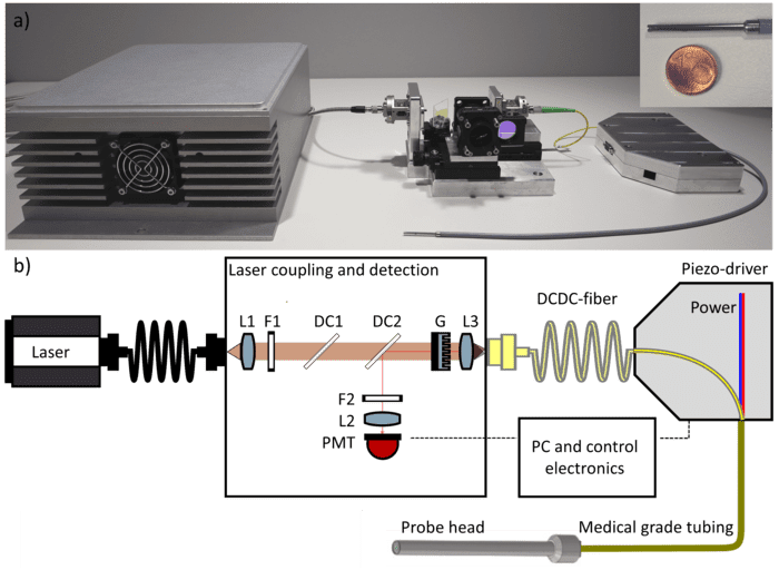
Featured Image: a) Photo of the endomicroscopic fiber probe and laser coupling unit. A picture of the stainless-steel shielded probe head of 2.4 mm diameter and a length of 39 mm is shown in the upper right corner in comparison with the size of one Euro cent coin. The optical fiber and the four connection cables of the piezo-scanner are guided in a 1 mm tube, which is enclosed in a sealed medical grade endoscopic tube of 4.5 mm in diameter. b) The fiber laser (AFS, Germany) is collimated by the lens L1, filtered from FWM at the CARS wavelength by F1 (750 nm long pass). The laser power is adjusted using a 1050 nm short pass dichroic mirror (DC1). A linear diffractive grating G of 39.4 μm grating period and lens L3 (f = 4 mm) couple pump and Stokes into the two different cores. Sample signals are guided back through the DCDC-fiber and detected by the PMT-detector after deflection at the dichroic mirror DC2. The bandpass filter F2 is selecting the non-linear signal of interest (CARS/SHG/TPEF) and the lens L2 focusing the signals onto the photo multiplier tube (PMT). Photo/Illustration: Ekaterina Pshenay-Severin, Hyeonsoo Bae, Karl Reichwald, Gregor Matz, Jörg Bierlich, Jens Kobelke, Adrian Lorenz, Anka Schwuchow, Tobias Meyer-Zedler, Michael Schmitt, Bernhard Messerschmidt, Juergen Popp

