Summary:
Researchers at Aarhus University have developed PathoPlex, a powerful and freely available tissue analysis method that uses multiplex imaging and machine learning to map over 100 proteins simultaneously, offering new insights into disease processes and drug responses.
Takeaways:
- Early Detection Potential: PathoPlex can detect early, otherwise invisible tissue changes in diseases like diabetes, enabling earlier diagnosis.
- Therapeutic Insight: The technique reveals how drugs, like SGLT2 inhibitors, impact disease processes directly in tissue, potentially guiding more personalized treatments.
- Open-Access Innovation: The system is freely available and adaptable, complete with a Python toolkit, making it accessible to labs worldwide for a wide range of disease research.
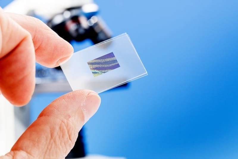
Researchers from Aarhus University, in a major international collaboration, have developed a groundbreaking method that can provide more information from the tissue samples doctors take from patients every day.
The new technique, called Pathology-oriented multiPlexing or PathoPlex, can look under a microscope at over 100 different proteins in the same small piece of tissue, instead of just 1-2 at a time, as is done now.
The technology, which has just been published in the prestigious journal Nature, combines advanced image processing with machine learning to map complex disease processes in detail.
“PathoPlex provides unique opportunities to explore the complex nature of human disease, which could have a direct impact on patient care,” explains Professor Victor Puelles from the Department of Clinical Medicine at Aarhus University.
Tissue Samples Allow for Early Disease Detection
In the study, researchers tested the technology on, among other things, specimens from patients with diabetes. Here they could detect complex disease processes that cannot be seen with conventional methods.
“We could see how diabetes affects the kidneys through an entire network of simultaneous changes,” says Victor Puelles.
Particularly noteworthy was that researchers could find changes in young diabetic patients before there were clear signs of kidney disease.
Medicine Effects Can be Better Measured
One of the most promising applications is the technology’s ability to measure how medicine works directly in tissue. The researchers examined the effect of SGLT2 inhibitors, a common type of diabetes medication, and found surprising results.
“We could see that treatment with SGLT2 inhibitors helped with some of the alterations linked to diabetes, but not all of them. While there is much more work to be done, our results raise important questions, for example, whether diabetic patients might need additional therapies to protect their kidneys,” says Victor Puelles.
PathoPlex: Free and Available to Everyone
Unlike many other research breakthroughs, the team has chosen to make their technology freely available. They have also made it flexible – with different solutions for small and large experiments, one of which uses a simple 3D printer as a liquid handling system. The researchers also include a step-by-step computational guide to perform image analysis, with a Python package called “spatiomic” available to the community.
“It was important for us to provide an open solution that the entire research community could use,” explains Victor Puelles.
Technology Can Have Applications to Many Diseases
Although the study focused on kidney diseases, researchers believe the technology can be used for almost all diseases where doctors need to examine tissues. For example, the team showed that PathoPlex works in liver and brain tissue, demonstrating how versatile it can be.
Before the technology can be used in hospitals, however, more work is needed. The researchers are working on simple solutions to fully automate the method, improvements to current benchmarking strategies, and concrete clinical applications that can show direct added value for patients.
Featured Image: Luchschen | Dreamstime.com


thank you for making this article very useful and keep up the good work