 Increasing threat to our health care system
By Craig Foreback, PhD
The statistics on the diabetes epidemic are staggering. Each November—American Diabetes Month—is an appropriate time to focus on the looming crisis faced by our health care system. According to data compiled by the Centers for Disease Control and Prevention (CDC)1 and the American Diabetes Association (ADA),2 8.3% of the US population has diabetes.
There are approximately one million type 1 diabetics and 18.8 million type 2 diabetics. It is estimated that there are an additional seven million undiagnosed diabetics. Diabetes is the leading cause of blindness and nontraumatic amputations, and it accounts for 43.8% of kidney failure. Direct and indirect medical expenditures totaled $174 billion—a 32% increase in the last 5 years. Diabetics have 2.3 times greater health care costs per capita, and one in 10 health care dollars is spent on the direct costs of treating the diabetic patient, and accounts for 30% of the total Medicare budget.
Figure 1 shows the dramatic increase in the number and percentage of the US population from 1958 to 2009. A major concern is the dramatic increase in type 2 diabetes especially in teens and children, largely attributable to the increase in obesity in these age groups. Melissa Merideth, MD, an endocrinologist at the University of Wisconsin Hospital and Clinics, Madison, Wis, has seen type 2 diabetes in patients as young as 7 years old. Risk factors for prediabetes and diabetes mellitus are shown in Table 1. The impact of diabetes on the future of the US health care system requires that laboratories and physicians work together to provide reliable and appropriate testing to screen and diagnose diabetes and monitor patients who are being treated for the disease.
SCREENING AND DIAGNOSIS OF DIABETES MELLITUS
The ADA has recently published standards of care for the screening, prevention, diagnosis, and management of diabetes3 (Table 2). There are issues with each of the recommended screening tests. While the majority of glucose testing performed on routine chemistry analyzers has been harmonized, the same cannot be said for point-of-care glucose meters. If screening is done by measuring glucose either fasting or with an oral glucose tolerance test (OGTT), the analysis needs to be conducted on a core laboratory analyzer. James Nichols, PhD, professor of pathology, microbiology, and immunology, Vanderbilt University School of Medicine, Nashville, Tenn, is opposed to the use of most glucose meters because they are just too imprecise to use for diagnosing diabetes. Many physicians want to use glucose meters to perform OGTT. This subject came up as a discussion on the American Association for Clinical Chemistry (AACC) General Chemistry listserv during the month of August. As might be expected, there was unanimous opposition to the practice.
The fasting requirement for the fasting plasma glucose (FPG) test is an issue since patients do not always comply, and some patients cannot tolerate the 75 g glucose load or the 3-day, high-carbohydrate diet required for the OGTT. The ADA has recommended the adoption of hemoglobin A1c (HgbA1c) for screening for diabetes. Nichols concurs with the recommendation since it does not require the patient to fast, but again opposes the use of point-of-care testing for the assay. A recent paper in Clinical Chemistry4 supports this position. An evaluation of eight HgbA1c point-of-care instruments concluded that six of the eight do not meet accepted performance criteria. Gary Tufel’s August 2012 article for CLP, based on an interview with Douglas Borses, Diazyme, Poway, Calif, summarizes the pros and cons of using HgbA1c as a screening test for diabetes.5
Most physicians now assume that all HgbA1c methods are equal. It is true that while the National Glycohemoglobin Standardization Program has dramatically improved conformance among assays, one must know the measurement principle behind the assay that is being used. Methods include ion-exchange, affinity, and immunoassay. Each of these methods can react differently with any individual sample. A discussion of the measurement principles can be found at Westgard QC.6 It is important to note that the use of HgbA1c is not universally accepted. In its practice guidelines, the AACC has recommended use of HgbA1c for screening; however, the American College of Endocrinology and the World Health Organization (WHO) say that endorsement is premature. Other limitations include the fact that Medicare will not reimburse for HgbA1c tests used for screening, and they cannot be used for the diagnosis of gestational diabetes.
The ADA criteria for diagnosing diabetes includes any one of the following:
HbgA1c >/= 6.5% using a standardized assay (using a NGSP-certified method)
FPG >/= 126 mg/dL (no caloric intake for
8 hours)
2-hour glucose >/= 200 during 75-gm OGTT
Classic symptoms of hyperglycemia and a random glucose > 200 mg/dL.
|
Table 1: Risk Factors for Prediabetes and Diabetes Mellitus • Genetic/familial |
MONITORING THE DIABETIC PATIENT
Traditionally, plasma glucose has been used to monitor diabetic patients, but the Diabetes Control and Complications Trial (DCCT) demonstrated that a new marker for chronic hyperglycemia was now available. The DCCT was a major clinical study conducted from 1983 to 1993 and funded by the National Institute of Diabetes and Digestive and Kidney Diseases. The study showed that keeping blood glucose levels as close to normal as possible slows the onset and progression of the eye, kidney, and nerve damage caused by diabetes. In fact, it demonstrated that any sustained lowering of blood glucose improves outcomes, even if the person has a history of poor control.
The study compared the effects of standard control of blood glucose versus intensive control on the complications of diabetes. Intensive control meant keeping HgbA1c levels as close as possible to the normal value of 6% or less. The HgbA1c blood test reflects a person’s average blood glucose over the last 2 to 3 months. The use of HgbA1c is universally accepted for long-term monitoring of diabetic patients and is used in conjunction with plasma or finger-stick glucose testing. However, many of the limitations of HgbA1c discussed in the diagnosis section also apply when using it as a monitor. An alternative test, discussed in Tufel’s article, overcomes some of the problems. Glycated albumin is particularly useful in the gestational diabetic and in patients with accelerated red cell turnover and anemia, since it is a short-term marker and is not affected by hemoglobin concentration.
GLUCOSE METERS
Portable meters account for the majority of the glucose determinations performed in the United States. Measurement of blood glucose concentrations are used in three major settings: (a) in acute- and chronic care facilities, including intensive care units (ICUs); (b) in physician’s offices; and (c) by patients at home, work, and school. Measurement in the last setting is termed self-monitoring of blood glucose (SMBG).
There has been intense interest in noninvasive glucose monitoring for obvious reasons. No noninvasive sensing technology is currently approved for clinical glucose measurements. Major technological hurdles must be overcome before noninvasive sensing technology will be sufficiently reliable to replace existing portable meters, implantable biosensors, or minimally invasive technologies. One company, Echo Therapeutics Inc, Philadelphia, is developing its needle-free system as a noninvasive, wireless, transdermal, continuous glucose monitoring system coupled to a device for transdermal drug administration. Results from a clinical trial of the system in general surgery and cardiothoracic surgery patients shows its promise as a continuous monitoring system that can potentially reduce errors, it requires less labor, and minimizes caregiver exposure to patient blood.
Figure 1: [Click here to view a larger image]
DIAGNOSIS AND MONITORING OF DIABETIC KETOACIDOSIS
The onset of ketosis requires changes in both adipose tissue metabolism and liver function. The primary substrates for ketone body formation are fatty acids from adipose stores. Ketone bodies are produced by the liver and used peripherally as an energy source when glucose is not readily available. Acetoacetate (AcAc) and 3-b-Hydroxybutyrate (bHB) are the two main ketone bodies; acetone is the third and least abundant. Strictly speaking, bHB is not chemically a ketone.
Ketone bodies are always present in the blood, and their levels increase during fasting and prolonged exercise. Diabetes is the most common pathological cause of elevated ketones. Diabetic ketoacidosis (DKA) occurs in poorly controlled diabetes, when low insulin concentrations and high levels of counter regulatory hormones initiate a cascade of events that increases plasma fatty acids and enhances fatty acid oxidation in the liver. The combination of increased hepatic ketone synthesis and decreased peripheral tissue metabolism leads to acetoacetate accumulation in the blood. A small fraction undergoes spontaneous decarboxylation to form acetone, but the majority is converted to bHB. In acute DKA, the ketone body ratio (bHB:AcAc) rises from normal (1:1) to as high as 10:1. In ketoacidosis, bHB levels are typically 78% to 80% of total ketone bodies.
The classic method for the detection and determination of ketones utilizes nitroprusside. The nitroprusside test is 10 times more sensitive to acetoacetic acid than to acetone and gives no reaction at all with bHB because it lacks a ketone ring. The fact that most of the commonly used methods for the determination of ketones measure only acetoacetate can produce a paradoxical situation. In a patient initially presenting in ketoacidosis the ketones, as measured by nitroprusside reaction, may be only weakly positive, whereas they are, in fact, high. This occurs because after initial treatment, bHB is converted to acetoacetate, which indicates a worsening condition.
DETERMINATION OF KETONE BODIES IN BODY FLUIDS
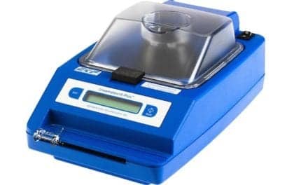
Quantitative enzymatic assays for the determination of BHB have been available for many years. A report from the University of Wisconsin Hospital and Clinics describes the development of an optimized method for the determination of serum bHB for use on a Cobas FARA.6 Umpierrez7 reported on the clinical utility determined by a reflectance meter in the management of DKA in 1995. The instrument was developed commercially as a benchtop analyzer (Ketosite; StanBio Laboratory, Boerne, Tex). The test uses a simple slide and requires 20 µL of serum. StanBio also supplies a liquid reagent with applications for numerous core lab analyzers.
The comparison of the determination of bHB versus nitroprusside (Acetest or Ketostix) is shown below:
bHB
• Quantitative
• Measures largest component of ketones
• Best and earliest indicator of ketosis
• Best predictor of resolution of ketoacidosis
Acetest
• Only measures acetoacetic acid and acetone (weakly)
• Qualitative assay often requiring multiple manual dilutions
• Increase lags behind symptoms of ketosis
• Decrease begins 2 to 4 hours after resolution of ketoacidosis
Several reports conclude that the use of Acetest may be misleading and should be avoided because the fall of acetoacetate lags behind the resolution of ketoacidosis. bHB levels correlate better with acid-base changes during the course of treatment for DKA than changes in acetoacetate concentrations. The study by Umpierrez7 highlights that all patients with bHB levels of 1.1 mmol/L or less had resolved their ketosis. In contrast, eight out of 15 patients in whom ketosis had been resolved, as demonstrated by blood gas and acid-base parameters, still had positive Acetest results for up to 24 hours after resolution. Similar results were obtained at Henry Ford Hospital, Detroit.8 In that study, bHB levels were just as sensitive as blood pH for demonstrating resolution of ketoacidosis.
Rapid determinations of bHB levels were useful in establishing the diagnosis of DKA and in the management of patients with prolonged metabolic acidosis, combined diabetic and lactic acidosis, and other mixed acid-base disorders. Direct measurement of bHB in serum improved laboratory turnaround time and replaced a subjective, qualitative result with a method that is quantitative and less subject to observer bias.9 In addition, the nitroprusside method has demonstrated susceptibility to false positive results from drugs containing free-sulfhydryl groups and false negative results from reagent deterioration.10,11 Improved clinical outcomes and enhanced cost efficiency have been attributed to blood testing of bHB.8 Improvements were seen in the following areas:
• Earlier detection of clinically significant ketosis
• Improved turnaround times
• Significant reduction in laboratory testing
• Faster resolution of ketoacidosis with significant reduction in the patients’ length of stay in the clinical decision unit.
Alternatives are now available for bHB in testing on multichannel chemistry analyzers and several point-of-care devices. Assays for bHB in blood are superior to Acetest or Ketostix in a number of respects. Physicians and medical facilities should no longer be performing nitroprusside-based testing to diagnose or monitor ketoacidosis.
In conclusion, it is imperative that accurate, precise, timely, and up-to-date testing be available for the screening, diagnosing, and monitoring of diabetes. Our health care system depends on it. n
Craig Foreback, PhD, is a contributing writer for CLP. For more information, contact Editor Judy O’Rourke, [email protected]
References
1. http://www.cdc.gov/diabetes/pubs/estimates.htm
2. www.diabetes.org
3. Standards of Medical Care. Diabetes Care. 2010;33:sup 1.
4. Lenters-Westra E, Slingerland R. Six of eight hemoglobin A1c point of care instruments do not meet the general accepted analytical performance criteria. Clin Chem. 2010;56(1):44-52.
5. Tufel G. HbA1c for Diabetes Testing Not Yet Universal. Clinical Lab Products. August 2012:33-35.
6. http://www.westgard.com/hba1c-methods-part5.htm
7. Koch DD, Feldbruegge DH. Optimized kinetic method for automated determination of b-hydroxybutyrate. Clin Chem. 1987;33(10):1761-1766.
8. Umpierrez G, Watts N, Phillips L. Clinical utility of ß-hydroxybutyrate determined by reflectance meter in the management of diabetic ketoacidosis. Diabetes Care. 1995;18(1):137-138.
9. Foreback C, Former Director of Clinical Chemistry, Henry Ford Hospital, Detroit, MI, White Paper, Clinical effectiveness of b-hydroxybutryate assays in a clinical decision
unit; 1998.
10. Foreback C. Beta-hydroxybutyrate and acetoacetic acid levels. Am J Clin Pathol. 1997;602-604.
11. Csako G. Unrecognized false–positive ketones from drugs containing free-sulfhydryl groups. JAMA. 1993;269(13):1364.
12. Csako G. False-positive results for ketones with the drug mesna and other free-sulfhydryl compounds. Clin Chem. 1987;33:289-292.
13. Goldstein D, Little R, Lorenz R, et al. Tests of glycemia in diabetes, Technical Review. Diabetes Care. 1995;18(6):896-90


