
Mastering tumor heterogeneity to unlock personalized medicine
By Mark C. Connelly, PhD
In solid tumor oncology, tissue biopsy is the gold standard for diagnosis, disease classification, and often therapy eligibility or selection. However, the continuing development of personalized medicine strategies has brought to light several limitations of tissue biopsies. For personalized medicine to be maximally effective, access to the current state of a tumor must be continuous and comprehensive.
Obtaining tissue biopsies can be difficult—particularly from patients with metastatic tumors—because the tumors may be small, present in multiple sites, or located in surgically high-risk or inaccessible locations. Even when a biopsy is feasible, it typically requires an invasive procedure, and samples only a limited region of a single tumor site. Finally, because repeated biopsies are unlikely, the single snapshot of a tumor provided by a tissue biopsy is neither continuous nor comprehensive. In short, tissue biopsies often do not provide the information required to deliver effective, tailored therapies against a disease that is constantly adapting and changing. In particular, tissue biopsies do little to facilitate an understanding of ‘tumor heterogeneity’—that is, the genetic diversity within a tumor. Considering that just one needle biopsy or surgical biopsy is unlikely to capture the complete genomic landscape of a particular cancer, many researchers are exploring the potential of liquid biopsies in an effort to better understand tumor heterogeneity and the role it plays in disease progression.1
A liquid biopsy is a simple, noninvasive procedure that uses a peripheral blood draw instead of tissue to access tumor-derived components. With results available in as little as a week, liquid biopsies facilitate near real-time monitoring of tumor phenotypic and genetic heterogeneity, which can enable clinicians and researchers to stay ahead of this ever-changing disease. This article explores several emerging liquid biopsy tools and research applications that provide more information from a wider variety of cell populations, which will hopefully lead to a better understanding of tumor heterogeneity. Such advances in technology are opening new areas of exploration and answering critical questions in pharmaceutical and clinical research that may soon be applicable in clinical settings, with the ultimate goal of unlocking a greater number of personalized therapeutic alternatives.
Mastering Tumor Heterogeneity and Clonal Tides
Designing better, more-targeted cancer therapies depends on a thorough understanding of the heterogeneity of a patient’s tumor and how it changes over time. Such heterogeneity arises from the different genetic mutations and dedifferentiations that exist within an individual tumor. While tumor cells are originally clonal, mutations create subpopulations of cells as the tumor grows.1
The development of such subpopulations has a profound effect on the phenotypes and genotypes of cells within a tumor, including how the subpopulations respond to various treatments. Some clones may initially be destroyed by a therapy, while others may thrive. Such an ebb and flow of ‘clonal tides’ throughout the course of the disease creates a constantly evolving landscape in which the cancer that remains after several rounds of treatment may have very different properties from the cancer that was present just a few months before. Frequent, regular, and noninvasive monitoring of tumor dynamics is necessary to rapidly identify clinical responses, resistance, and changes in clonal dominance over the course of therapy. Such monitoring will ultimately aid in the development, choice, combination, and sequence of personalized therapies, much as it does today in the treatment of HIV. Combined targeted therapies that attack all or most of the presenting variants may prove to be more effective at achieving sustained responses. Personally tailored sequential or pulsed therapies—with optimized administration guided by liquid biopsy—could prolong or sustain remission while minimizing toxicities.
Realizing this vision will require extensive clinical research, as well as a deep understanding of the biology and drug sensitivity associated with various genetic subpopulations. In order to obtain this information, researchers need the ability to study all classes of tumor-associated analytes (TAAs), including access to technology that preserves and isolates all TAA classes conveniently, accurately, and reproducibly across laboratories.
Getting a Detailed Picture
Researchers can use liquid biopsies to enumerate and analyze various classes of TAAs, each of which provides different information about the current state of the tumor. Classes include circulating tumor cells (CTCs), circulating tumor DNA (ctDNA) from plasma, extracellular vesicles (EVs), and exosomes (EXOs). Tapping into each class of TAA may provide a comprehensive and detailed picture of a patient’s disease state, with potentially clinically actionable information. Although CTCs were first identified as early as 1869, automated and validated methods for their isolation and analysis have only become available in the past 2 decades.
CTCs have been shown to be clinically useful as predictors of overall survival and progression-free survival in patients with metastatic breast, colorectal, or prostate cancer.2 The cells can be used to examine current CTC phenotypes and identify which patients are at highest risk of disease progression. CTCs can also be an excellent source of tumor DNA and RNA, essentially free of genomic nucleic acid contamination, for sequencing and mutation analysis. Recently, researchers have begun using ctDNA found in plasma to probe various cancers, such as pancreatic and central nervous system tumors, or to monitor patients with lung cancer and develop mutation-specific treatment options.3
While not able to form distant metastases, the relatively abundant EVs are anuclear cell fragments that are beginning to unlock information on the current status of the tumor microenvironment. Exosomes are small packets (~200 nM) shed by cells; they can transmit messages, and even change gene expression in distant targeted cells. Understanding the language of exosomes could provide important signals that may telegraph the tumor’s next move. To be broadly applicable, this kind of holistic information must be accessible to all researchers and practitioners, not just those at large academic centers. Liquid biopsies are well suited to meet this need, because a tube of blood can be drawn at any location and sent to whatever laboratory is performing the necessary analyses.
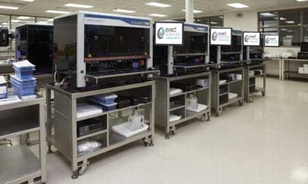
The proprietary CellSave preservative tube, part of the CellSearch CTC system from Menarini Silicon Biosystems, Huntingdon Valley, Pa, stabilizes and preserves CTCs, ctDNA, EVs, and EXOs for up to 96 hours for transport to the laboratory (Figure 1). However, it is critical that researchers take steps to preserve the full range of TAAs during transport to the laboratory. CTCs, RNA, and ctDNA are fragile and easily damaged during shipping. While several commercially available solutions can preserve ctDNA, most will actively destroy CTCs, EVs, and EXOs in the process. Other solutions can protect most CTCs, DNA (both nuclear and ctDNA), EVs, and EXOs, but will severely damage any RNA found within any class of TAAs. CellRescue is a recent advance in cellular preservation technology from Menarini Silicon Biosystems. Currently available for research use only, CellRescue holds the promise of preserving all classes of TAA found in a liquid biopsy, including target-cell RNA, in a single tube. Preserving the full range of TAAs in this way would greatly reduce the type and number of sample tubes to be drawn, decrease the amount of blood required for a comprehensive TAA analysis, and maximize the utility of liquid biopsies for facilitating a better understanding of the disease.
A More Complete Liquid Biopsy
In the past two decades, liquid biopsy researchers have established that CTCs are more likely to represent the full genetic heterogeneity of a tumor compared to a tissue biopsy, which takes a sample from a limited region of a single tumor site. By contrast, CTCs can come from multiple sites in a single patient, or even multiple sites within a single tumor mass. In addition, the clinical significance of these cells has been demonstrated by the strong association between the number of CTCs and changes in overall and progression-free survival in breast, colorectal, and prostate cancer.
Evidence of the clinical significance of CTCs has been firmly established for CTCs isolated and enumerated using the CellSearch CTC system from Menarini Silicon Biosystems. The system is currently the only in vitro diagnostic device cleared by FDA for enumerating CTCs. As few as 3 to 5 CTCs present in 7.5 mL of blood can predict a dramatic change in a patient’s prognosis. Having the capability to detect this numeric change may be useful for monitoring patients.2 However, the number of CTCs alone does not provide insight into their heterogeneity. To effectively monitor tumor heterogeneity using these rare cells, they must be enriched and separated into single cells, so that the mutations within each of the cells can be examined. This technique enables researchers to define the clonal subsets that are present, and study the specific molecular changes that occur in the tumor over time.
The DEPArray technology from Menarini Silicon Biosystems is an image-based cell-sorting technology that combines microelectronics and microfluidics in an automated platform to isolate pure, single cells from a mixed cellular sample. While CellSearch can enrich, identify, and enumerate CTCs, the DEPArray technology can isolate and harvest individual tumor cells, or pools of tumor cells, with 100% purity. The DEPArray technology is currently available for research use only (Figure 2). Harvested cells can be subjected to single-cell molecular characterization, ranging from chromosome numerical aberration studies to complete genomic sequence analysis.
The ability to analyze mutations, define a tumor’s heterogeneity, and follow the changes in mutation patterns in near real time will hopefully lead to the development of new and more-effective personalized treatment regimens. Using this technology, researchers will soon be able to obtain even more information from a wider variety of cell populations. While the current DEPArray platform offers up to six optical channels to identify and analyze CTCs of interest, forthcoming technology will increase this capacity to nine optical channels. Offering more fluorescence channels to examine the heterogeneity of marker expression on various rare cells will enable researchers to simultaneously identify, characterize, and harvest multiple cell populations. Such different cell populations, expressing diverse markers of interest, may be found to represent varied pathologic and clinical consequences for the patient.
Not Just Data: AI Delivers More Information
As technology increases the ability of researchers to measure a greater number of TAAs—each of which may be associated with multiple qualitative and quantitative test results—the available data rapidly become multidimensional to the point of being incomprehensible. Artificial intelligence (AI) has shown the potential to assist researchers with uncovering associations, characteristics, and parameters previously hidden within the data; as well as with processing all TAA data from a given patient sample and turning it into intelligible, actionable information.
Researchers currently identify CTCs and EVs by manually reviewing images. But an automated, AI-driven process is being constructed that will bring greater ease and consistency to CTC analysis (Figure 3). In a recent study, the use of deep convolutional neural networks to classify cells obtained by CellSearch technology automatically identified and characterized CTCs from metastatic cancer and benign diseases with an accuracy, sensitivity, and specificity of over 96%. In addition to obtaining CTC counts that were predictive of overall survival, this automated quantification identified novel features that defined subclasses of cells previously unrecognized by skilled manual reviewers.4
The application of AI to CTCs and other TAAs obtained through liquid biopsy has the potential to provide researchers with a deeper understanding of the complex pathophysiology of cancer, while at the same time making that complexity easier to comprehend and easier to visualize. Transforming such highly complex data into clinically useful information could improve patient monitoring, uncover new biomarkers, and help to optimize treatment selection, timing, and delivery.
Early Evaluation of Responses to Therapy
During the process of developing new pharmaceuticals or combination therapies for cancer treatment, it is critical to know whether patients are responding favorably to an experimental therapy as soon as possible. In addition to such classic outcome indicators as progression-free survival and overall survival, imaging is commonly used as an in-process means of determining whether there has been a reduction in tumor size or in the number of metastatic sites. Yet, several months can go by before the size or number of tumors show meaningful changes. Likewise, progression-free survival and overall survival can take months, or even years, before a statistically significant change can be observed.
In order to accelerate clinical evaluations and decrease the time required to get promising new therapies to the clinic, researchers need biomarkers that can reliably indicate whether patients are responding favorably soon after the start of treatment. Researchers working with metastatic castration-resistant prostate cancer (mCRPC) do not yet have such a reliable early-response biomarker. Clinical trials for mCRPC almost always measure a patient’s level of prostate-specific antigen (PSA), even though PSA is not considered to be a reliable early indicator of treatment response. In fact, no drugs have been approved based solely on their ability to elicit a favorable change in PSA levels.5
However, new research supports the potential use of CTCs as early response biomarkers for mCRPC. An analysis of more than 3,000 patients across five Phase III randomized controlled trials indicates that using CTC counts obtained from a liquid biopsy can provide a reliable early assessment of response to treatment.6 The researchers concluded that a change in the number of CTCs at baseline, and then after 12 weeks of therapy, was the best indicator of treatment response. In addition, they reported that a reduction in CTC count was the best predictor of improved patient outcomes—independent of either PSA levels or changes in PSA levels. Menarini Silicon Biosystems is working with FDA to qualify CTCs as an early response biomarker for use in mCRPC clinical trials. If qualified by the agency, the use of CTCs from a liquid biopsy as a biomarker would have the potential to accelerate the drug development process. A reliable early biomarker would make it possible to eliminate ineffective treatment candidates sooner, and would also accelerate the development of beneficial pharmaceutical candidates in the fight against mCRPC.
New Frontiers for Liquid Biopsy and New Ways to Conduct Clinical Research
The CellSearch and DEPArray workflows are currently being used in research to extend the utility of liquid biopsy in several areas of cancer, including melanoma and multiple myeloma (see Sidebar). Multiple myeloma is the second most common hematological cancer in the United States, and it remains incurable, with a 5-year survival rate of only 45%.7 Because multiple myeloma is a bone marrow cancer, diagnosis of the disease requires an invasive bone marrow biopsy. However, myeloma cells also transit from the bone marrow to the bloodstream, so detection and characterization of myeloma cells from peripheral blood samples have long been used to monitor the disease.
Researchers currently rely on flow cytometry for the detection of circulating multiple myeloma cells. However, flow cytometry is limited by a lack of standardization across laboratories, and a lack of sensitivity for detecting low numbers of rare circulating multiple myeloma cells—particularly when monitoring minimal residual disease. The immunomagnetic enrichment of circulating multiple myeloma cells from blood affords much greater sensitivity than flow cytometry because the technology specifically captures the multiple myeloma cells while removing most other white blood cells. Enriching circulating multiple myeloma cells more than a thousandfold over other blood cells means larger volumes of blood can be analyzed compared to flow cytometry. A recent study used the CellSearch system to capture and detect the number of circulating multiple myeloma cells obtained from liquid biopsies, enabling the researchers to monitor the number of cells present at baseline, detected during therapy and remission, and present at recurrence.7
The DEPArray technology can be used to further purify and harvest individual cells for detailed single-cell or pooled molecular analysis. Studying cells for both phenotypic and molecular changes has revealed the simultaneous presence of multiple molecular subclones of circulating multiple myeloma cells in a patient (Figure 4). Although more research is needed to investigate how molecular subclones change over time and in response to therapy, the ability to observe these specific molecular changes in real time will likely have therapeutic implications for such an aggressive and dynamic cancer. Studies like this are shedding light on how to control the clonal tides that are now being recognized in this and other cancers. The combined liquid biopsy workflow of CellSearch and DEPArray enables researchers to investigate premyeloma, smoldering myeloma, and multiple myeloma in new and exciting ways—without having to perform invasive bone marrow biopsies. The global laboratory services unit of Menarini Silicon Biosystems currently offers a CLIA-registered laboratory-developed test for circulating multiple myeloma cells, with plans to release a research use only commercial kit in late 2020.
Unlocking Personalized Medicine
As a result of tumor heterogeneity, solid tumor samples taken years or even months ago cannot provide researchers with a temporally accurate understanding of the disease in its current molecular state. Fortunately, liquid biopsy tools and applications are moving researchers closer to being able to examine tumor subpopulations and the impact of clonal tides on disease progression in real-time through noninvasive means. Such liquid biopsy workflows can provide deep insights and expansive information about a tumor’s current status. When combined with AI, researchers will be able to convert all that data into biologically and clinically relevant information. Applying liquid biopsy to disease management has the potential to unlock personalized medicine by providing researchers with current information regarding tumor dynamics, helping them stay one step ahead of an ever-changing disease.
Mark C. Connelly, PhD, is the chief research and development officer at Menarini Silicon Biosystems. He has been conducting research into circulating tumor cells and developing liquid biopsy technologies for 18 years. The DEPArray system technology, CellRescue tubes, and circulating multiple myeloma cell kit described here are currently available for research use only, and are not for use in diagnostic procedures. The performance characteristics, safety, and effectiveness of the products have not been established and are not cleared or approved by FDA.
REFERENCES
1. Gerlinger M, Rowan AJ, Horswell S, et al. Intratumor heterogeneity and branched evolution revealed by multiregion sequencing. N Engl J Med. 2012;366(10):883–892; doi: 10.1056/nejmoa1113205.
2. CellSearch circulating tumor cell kit (epithelial) [e631600006_EN LBL-0018, instructions for use, online]. Huntingdon Valley, Pa: Menarini Silicon Biosystems, 2017. Available at: https://documents.cellsearchctc.com/pdf/e631600006/e631600006_EN.pdf. Accessed June 9, 2020.
3. Normanno N, Denis MG, Thress KS, Ratcliffe M, Reck M. Guide to detecting epidermal growth factor receptor (EGFR) mutations in ctDNA of patients with advanced non-small cell lung cancer. Oncotarget. 2017;8(7):12501–12516; doi: 10.18632/oncotarget.13915.
4. Zeune LL, Boink YE, van Dalum G, et al. Deep learning of circulating tumor cells. Nature Machine Learning. 2020;2:124–133; doi: 10.1038/s4256-020-0153-x.
5. Scher HI, Morris MJ, Stadler WM, et al. Trial design and objectives for castration-resistant prostate cancer: updated recommendations from the prostate cancer clinical trials working group 3. J Clin Oncol. 2016;34(12):1402–1418; doi: 10.1200/jco.2015.64.2702.
6. Heller G, McCormack R, Kheoh T, et al. Circulating tumor cell number as a response measure of prolonged survival for metastatic castration-resistant prostate cancer: a comparison with prostate-specific antigen across five randomized phase III clinical trials. J Clin Oncol. 2018;36(6):572–580; doi: 10.1200/jco.2017.75.2998.
7. Foulk B, Schaffer M, Gross S, et al. Enumeration and characterization of circulating multiple myeloma cells in patients with plasma cell disorders. Br J Haematol. 2018;180(1):71–81; doi: 10.1111/bjh.15003.
Featured image: The proprietary CellSave preservative tube stabilizes and preserves tumor-associated analytes for up to 96 hours, during transport to the laboratory.
