By Matthew Wildrick
Summary:
Next-generation AI-powered digital pathology platforms are revolutionizing wet mount slide handling, enabling reliable, automated imaging of challenging specimens and unifying clinical and anatomic pathology workflows to improve efficiency and diagnostic accuracy.
Takeaways:
- Breakthrough in Wet Mount Digitization:
Modern systems overcome longstanding optical and time-sensitive challenges of wet mount slides—like variable focus, evaporation, and image degradation—through AI-driven automation and intelligent slide handling. - Workflow Standardization and Scalability:
These platforms support integrated, repeatable workflows across various specimen types and lab settings, reducing manual intervention and enabling scalable, cross-specialty deployments without extensive retraining or infrastructure investment. - Enhanced Diagnostic Speed and Accuracy:
Automated focus optimization, label recognition, and AI-powered quality control allow faster turnaround and consistent imaging, particularly for complex applications such as human fecal trichrome (HFT) analysis in microbiology labs.
In the fast-paced world of clinical laboratories, the handling of wet mount slides has long been a critical yet challenging aspect of diagnostic workflows. Traditional whole slide imaging (WSI) systems have struggled with liquid-based specimens and smears due to their sparse and non-uniform nature, creating bottlenecks in lab operations. However, next-generation solutions are breaking through these barriers, enabling comprehensive digitization of wet mount slides across multiple specialties.
Modern AI-powered platforms now offer specialized capabilities for handling wet mount slides, particularly in microbiology and parasitology applications. These solutions demonstrate how advanced systems can overcome the challenges of digitizing liquid-based specimens, including human fecal trichrome (HFT) use cases and other challenging sample types. The technology represents a significant advancement in unifying anatomic and clinical pathology workflows under a single digital platform.
These innovations are particularly valuable for medium- and high-throughput laboratories looking to create a more efficient, integrated approach to digital pathology, and handle the full spectrum of laboratory needs.
The Complex Nature of Wet Mount Slides
Wet mount slides present unique challenges that have historically made them particularly difficult to digitize. These challenges span multiple technical domains, including:
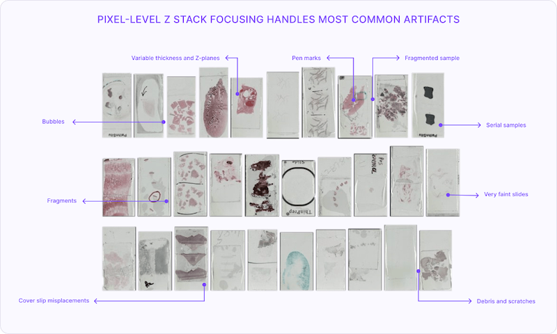
- Optical and Imaging Complexities: The very nature of wet mounts creates fundamental optical challenges. The interface between liquid and air creates complex light paths, making automated focus and image clarity significantly more difficult to achieve. Additionally, the dynamic nature of wet mounts means that cells or organisms can shift or float during scanning, leading to blurry or inconsistent images. If not handled properly, bubbles and debris in wet mount slides can interfere with AI detection algorithms and confuse tissue classifiers, if utilized as part of digital pathology initiatives. It is also imperative to design a WSI system that implements a stable placement of the wet mount slides in the scanner to avoid impacting specimens.
- Time-Sensitive Nature: Perhaps the most critical challenge is the time-sensitive nature of wet mounts. These specimens can only be viable for a few hours, making them incompatible with batch scanning approaches if not handled rapidly. The risk of evaporation can lead to specimen degradation mid-scan. Since wet mounts are typically unfixed, cell morphology changes rapidly, potentially affecting consistency in AI-based interpretation and making timing crucial for accurate results.
Limitations of Traditional Digital Pathology Scanners
For years, diagnostic labs have struggled with the inherent limitations of their conventional whole slide imaging scanners when it comes to wet mount slide handling.
These traditional systems typically assume static, fixed samples, but wet mounts may have variable depths that confuse autofocus systems. Non-uniform thickness due to liquid spread affects scanner calibration and often results in out-of-focus regions. Moreover, traditional WSI systems aren’t built for handling slides with loose liquids, creating risks of damage to the digital slide scanner or the specimen on the wet mount slides.
These systems also require technicians to simultaneously manage both the physical handling of slides manually in preparation for image scanning, and the scanning process, creating a complex dance of precision and timing. This approach does not result in a completely repeatable process workflow within lab operations, and introduces multiple points of potential error, from improper slide placement to inconsistent scanning parameters. Overall, with these challenges of traditional systems in place, return on investment can hardly be justified as lab operations scale to enable digitization.
Overall, given these limitations, labs face significant operational burden due to manual intervention requirements, and the resulting reduced throughput, for wet mount slides. Moreover, the transition from R&D efforts with digital pathology to full-scale clinical workflows becomes almost impossible without a repeatable process, since scaling up traditional systems often requires justification for significant staff training and infrastructure investment.
A New Path Forward
The innovative approach of automating the scanning process for wet mount slide handling addresses these challenges head-on. Today, it is possible to build fully autonomous scanner hardware that can dynamically adapt to the presence of different pathology slides and apply the appropriate whole slide imaging techniques in software.
Just as modern smartphones combine advanced camera hardware with intelligent software to automatically adjust focus, exposure, and color balance based on their environment, WSI systems can now leverage similar technological innovations. These mobile devices that we use in our daily lives use sophisticated AI algorithms to analyze the scene in real-time, optimize settings for different lighting conditions, and enhance image quality. When it comes to digital pathology scanners, utilizing latest innovations in agile software development and AI has proven to offer highest quality imaging for different types of slides and specimen characteristics.
By adopting such new technology principles, modern whole slide imaging systems can create repeatable workflows. By eliminating traditionally manual slide preparation and automating quality control in software, laboratories can achieve new levels of efficiency, especially in processing challenging specimens, such as HFT analysis. For instance, high-volume microbiology departments can significantly improve turnaround times and diagnostic accuracy when they can consistently digitize and analyze wet mount slides.
Modern whole slide imaging systems also incorporate intelligent automation that handles physical slide manipulation with remarkable precision while maintaining specimen integrity. This separation allows for optimized scanning parameters and consistent handling procedures, regardless of the volume of slides being processed. The system’s AI-driven imaging capabilities are further enhanced by advanced label recognition technology that automatically reads and processes slide information during scanning. As a result, the platform can not only identify specimen types from labels but also automatically adjust scanning parameters based on this information, ensuring optimal image quality for each sample.
These advanced capabilities, which are enabled with new generation of intelligent hardware and software solutions, help maintain consistent quality, especially when handling challenging wet mount slides. They allow laboratories to consolidate their workflows into a single, centralized operation under a standard set of rules and workflows, rather than maintaining separate digital systems across different locations and specialties. This strategic consolidation delivers significant time and cost savings by breaking down departmental silos, reducing redundant equipment investments, and optimizing resources.
Enabling the Digital Pathology Ecosystem
The impact of such new innovations extends to the broader digital pathology ecosystem with high-quality and consistent image capture creating a solid foundation for downstream analysis.
For example, in microbiology laboratories, modern systems can enable AI-powered analysis of human fecal trichrome (HFT) wet mount slides, automatically annotating potential anomalies. This intelligent screening process significantly reduces manual review time by directing pathologists to specific areas of interest only on positive slides, rather than requiring microscopic examination of every slide.
For such specialized applications, these modern systems can ensure proper focus and tissue coverage across any slide specimen, whether it’s a routine H&E or a challenging wet mount. This reduces the need for manual quality checks, allowing viewing software provided by the Image Management System (IMS) to focus on delivering advanced visualization features.
Built-in quality assurance capabilities can further enhance the delivered value by automatically detecting and flagging potential issues during the scanning process, only passing on validated high-quality images to AI algorithms. This proactive and automated quality control is particularly valuable for applications like HFT analysis, where it helps maintain the integrity of both the diagnostic process and AI training datasets.
Real-World Impact
With new modern digital pathology systems in place, laboratories report significant reduction in processing times and error rates, and improved clinical turnaround times. Standardization of slide handling workflows across different specimens leads to increased efficiency in resource utilization for equipment and staff.
It has been critical to easily scale the deployment of these systems, within the same lab or within labs across multiple locations, without breaking the bank. This has translated to faster adoption curves for organizations that take the solution from their R&D labs to production environments across clinical laboratories.
Looking Forward
As labs face increasing pressure to improve efficiency, laser focus on end-to-end lab workflows will be key. Instead of focusing on improving the handling of just one type of specimen and slide type with digital pathology, it is important for medical directors to demand the deployment of flexible systems that can adapt to the realities of the lab environment at any given day. These systems should provide the ability to automatically learn and scale as the real-time requirements change across clinical labs, without requiring new manual workflows.
These intelligent solutions should be designed to handle multiple specialties: microbiology slides for various organisms, anatomic pathology cases including tissue biopsies and frozen sections, cytopathology workflows such as liquid-based cytology and conventional Pap smears, as well as hematopathology specimens like bone marrow aspirates and peripheral smears.
This digital and automated future for pathology slide handling in laboratory diagnostics is not a good-to-have but a must-have requirement as pathologists continue to demand highest standards of accuracy and efficiency. This strategic approach to handle wet mount slides demonstrates how innovative thinking can transform traditional processes, creating new possibilities for laboratory operations and, ultimately, patient care.
About the Author
Matthew Wildrick is senior director of Solutions Architecture at Pramana.
Featured Image: One example in handling slides that are challenging in nature is the use of pixel-level fast focus sampling and dynamic calculation of the tissue’s z plane. Using this technology, modern systems can make in-line decisions based on tissue type, thickness and slide artifacts like annotations and debris. Image: Pramana


