
Starting with whole-slide imaging, a cascade of advances is waiting in the wings
By Russell Granzow

Russell Granzow, Philips Digital Pathology Solutions.
Slowly but surely, healthcare is going digital. Many of the recent innovations in healthcare, from telemedicine and smart devices to the growing capabilities in managing big data, can be traced back to the adoption of new digital technologies that have fostered a different way of working. Across medicine’s varied specialty areas, however, the adoption and progress of digital technology advances varies significantly.
In pathology, and particularly in molecular pathology, there are an increasing number of tests, and an increasing demand to support faster, more-accurate disease diagnosis—and subsequent treatment and care—particularly for cancer. But while the vast majority of cancer diagnoses are made or confirmed by pathologists, the demand for such skilled professionals is not currently being matched by a growing number of new pathology graduates. In fact, figures for the United States show that the number of active physicians in pathology decreased by 10.4% between 2008 and 2013, and 60.7% of active pathologists are 55 or older.1
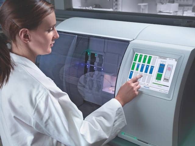
A lab technician initiates scanning with the IntelliSite ultra fast scanner.
Pathology laboratories are among the last healthcare environments to benefit from fully digitized workflows. In most hospitals and pathology laboratories today, tissue-based diagnoses are still rendered by pathologists using the traditional process of analyzing glass slide samples using a microscope. This traditional way of working comes with challenges—aside from the inherent delays built into its processes—including the time and risks associated with transferring physical glass slides. However, change is starting to emerge, with the introduction and adoption of digital pathology systems in both research and clinical settings.
JUSTIFYING THE TRANSITION
Personalized medicine and genomics offer the promise of individualizing healthcare—a particularly strong motivator in the field of oncology, where the demand for better and more-efficient care permeates the dialogue of healthcare professionals. Pathology will play a central role in this evolution. As the work of diagnosing and treating cancer becomes more complex, however, so too does the work of the pathologist and, by extension, that of the entire pathology department.
While clinicians in the specialty of pathology may be decreasing, oncologists are facing mounting pressure to curtail the pervasive disease at the center of their specialty. Statistics compiled by the International Agency for Research on Cancer project that new cancer cases will rise 70% over the next 2 decades, from 14 million in 2012 to 22 million per year in 2035.2
Even more troubling is the lack of concordance in the definitive diagnoses of cancer. A study from the American Society of Clinical Oncology found that for common HER2 breast cancer, approximately 20% of testing may be inaccurate—and this finding is for just one type of cancer.3 Another report issued by the UK’s National Patient Safety Agency found that protracted decisionmaking in pathology accounts for 41% of delays in cancer diagnosis, and is a leading cause of patient safety incidents.4
Pathologists are still just beginning to learn how to manage the massive amounts of rich imaging data they procure every day, but it is clear that there is room for improvement over existing processes. Now is the time when pathologists most need flexibility in harnessing the imaging data at their disposal, in order to put the process of definitive diagnosis on a path toward making personalized medicine a widespread reality.

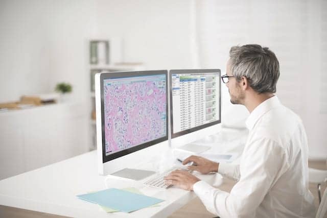
The case viewer of the IntelliSite pathologist suite.
The transition from microscope-based image analysis to digital pathology may seem like a natural progression, in line with the rising tide of widely varied digital technologies that are already being integrated into the practice of medicine. However, the adoption of digital technology systems has been slowed because of concerns over the potential for such systems to disrupt longstanding practices that are deeply embedded in the pathology profession. In addition, some skeptics continue to raise questions about the investments necessary to implement a digital pathology practice—not to mention the willingness of public and private payors to reimburse providers for making the transition, and the amount of time required for a practice to realize a meaningful return on its investments in such advanced technologies.
Additional obstacles arise from regulatory concerns, based in part on the difficulty of using traditional approaches in clinical studies to test or reproduce the skill of the microscope-aided human eye and compare it to the power of digital technologies. Consequently, the reproducibility and standardization associated with real-world use of digital pathology technologies have yet to be evaluated on a large scale.
Changes of any sort can be difficult, and often involve trial and some time to make adjustments. But experts remain certain that the inevitable digitization of pathology will lead to numerous benefits and new opportunities for pathology departments, patients, and the future potential of precision oncology.
DIGITAL PATHOLOGY SUPPORTS COLLABORATION AND EFFICIENCY
As more pathology departments move from traditional to digital workflows, many aspects of pathologists’ daily work are affected. Coincidently, but not surprisingly, such changes mirror those that occurred in radiology departments more than 2 decades ago, when advanced digital imaging technologies were first introduced into clinical practice.
Digital pathology promises a similar transformation for the clinical laboratory community, enabling labs to further improve the systematic management of pathology imaging data through enhanced data management, backup, storage, and archiving capabilities. Two important functions of digital pathology are to search and share pathology images and reports very quickly. By augmenting workflows with digital pathology solutions, advanced integration with other hospital information systems—such as electronic medical records, laboratory information management systems, and so on—can benefit pathologists by enabling them to search for patient pathology and related clinical information using more intuitive means, such as patient IDs or case IDs.
ADVANCING TECHNOLOGIES
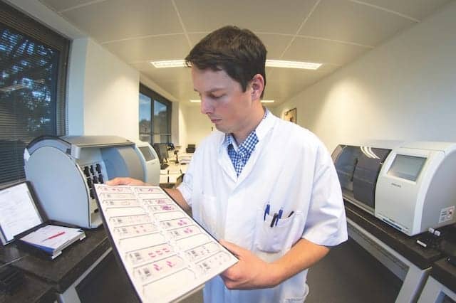
A lab technician at the Laboratory for Pathology East Netherlands Foundation (LabPON) reviews tissue slides for scanning.
In traditional pathology workflows, glass slide samples are prepared manually, examined under a microscope, and physically shipped for external consultation. This process is time-consuming and can be precarious, as there is a risk of losing or damaging the specimens in transit. By contrast, the latest and most promising imaging modality to disrupt the clinical lab sector is digital pathology whole-slide imaging, which entails the scanning of glass slides to produce precise digital image replicas in turn speeding and simplifying access to histopathology information across the organization.
Contributing to the advance of this field is the IntelliSite pathology solution by Philips, Best, the Netherlands, an automated system for creating and managing digital images of pathology specimens. The IntelliSite pathology solution is a highly scalable platform featuring three core elements:
- IntelliSite ultra fast scanner, a high-throughput bright-field slide scanner designed to accommodate current histopathology needs for routine use in high-volume labs and integrated pathology networks. The scanner has a storage capacity of 300 barcoded slides, which allows the scanner to run overnight. It also supports a continuous scan mode; it can be loaded and reloaded at any time without disrupting the scanning process.
- IntelliSite image management system works to improve the efficiency and effectiveness of pathology labs. The open and scalable design offers optimal integration with the lab’s workflow and IT infrastructure environment.
- IntelliSite pathologist suite, designed to enable pathologists to review cases as quickly as possible; provides easy access to information and resources to enable better-informed decision-making.
The system’s advanced software tools manage the scanning, storage, presentation and sharing of information.
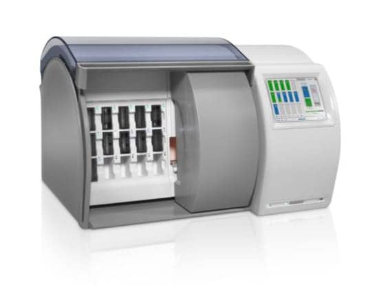
The IntelliSite ultra fast scanner by Philips Digital Pathology Solutions, Best, the Netherlands.
The IntelliSite pathology solution is CE marked and commercially available for in vitro diagnostic use under the European Union’s In Vitro Diagnostics Directive. The system is also licensed by Health Canada, and registered in Australia, Singapore, and the Middle East, for in vitro diagnostic use. In the United States, the system is indicated for in vitro diagnostic use for manual reading of the digital HER2 application.
In traditional pathology workflows, many steps in the specimen preparation process are performed manually, making it difficult to keep track of specimens and where they stand in the process. Using a digital pathology solution permits pathologists to identify and keep track of where specific specimens stand in the review process, and enables the department to determine whether schedules are following expected protocols, or why delays may be occurring.

In traditional pathology laboratories, many steps in the specimen preparation process are performed manually.
Among the advantages of a digital pathology solution is the fact that digital slide images can be more conveniently and safely distributed for external consultation, by being sent directly to the external consultant, or by giving the consultant access to view the images from a remote location.
Efficiencies in lab operations are another promising mark of digital pathology. Pathologists have traditionally performed diagnostic readings by viewing glass slides while standing (or sitting) over a microscope. An obvious but important element of pathology laboratory digitization is that it fundamentally changes the pathologist’s primary workstation, moving the pathologist from the microscope to a computer workstation. This change can help to ease the physical fatigue and strain placed on pathologists from leaning over a microscope for many hours a day.
The user screen of the IntelliSite pathologist suite permits side-by-side comparison of digital pathology images.
As pathologists acclimate to computer-based analysis, and vendors address opportunities related to the ergonomics of a digital image, the digital workstation offers several advantages when compared to microscopy alone. It is now possible to view different stains of a tissue section side-by-side, automatically aligned to allow for a more-direct comparison and, ultimately, outcomes that are more accurate. Additionally, digital workstations make image measurements easier, and also facilitate the retrieval and comparison of older images from the same patient.
As the field of pathology makes its digital transformation, including the use of advanced imaging analytics, it should be underscored that the aim of such transitions is not to replace the knowledge or experience of the pathologist. Instead, digital systems leverage imaging technologies and related computing capabilities to advance the work and collaboration of pathologists in and outside of the lab.

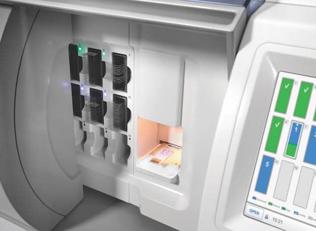
Closeup of a slide cartridge for the IntelliSite ultra fast scanner.
From the standpoint of workflow efficiency and collaboration, digital pathology offers clear benefits. But it is equally important—or maybe even more important—to consider the potential value of digital pathology in the larger context of newly emerging value-based healthcare systems. The worldwide incidence of cancer is continuing to grow, leading to corresponding increases in the workloads and pressures on pathology departments. Traditional pathology systems and protocols may no longer provide pathology laboratories with the tools and resources they will need to respond adequately to such rising demands.
In order to elevate their diagnostic processes to the next level, pathologists around the globe have started to adopt digital pathology tools. Nevertheless, digital pathology is far from being considered standard practice, and there is still a long road ahead before a majority of pathology laboratories will have made the transition—even in leading cancer centers.
Glass slide scanning in progress on the IntelliSite ultra fast scanner.
Major obstacles to converting pathology laboratories to a digital workflow include concerns over the levels of investment required to make the transition, and whether lab revenues can ever be sufficient to produce a reasonable return on such investments. For most lab organizations, cost-effectiveness is a key consideration; investments that are not fruitful can frequently become significant factors that inhibit innovation (For more information, see “The Business Case for Digital Pathology“). However, the financial benefits of digital pathology are gradually becoming more evident, as an increasing number of labs are recognizing that digital pathology can increase the revenue of a pathology lab and its hospital system, while also reducing costs through better lab utilization in networked hospital settings—including near elimination of costs for the transport and storage of glass slides. Indeed, as the benefits resulting from digital pathology are shared with other clinical stakeholders, such as oncologists and surgeons, return on investment calculations are beginning to support the transition.
THE ROAD AHEAD
The future of effective, value-based healthcare will be enabled by health technology solutions that can effectively manage the increasingly complex requirements of all stakeholders in the healthcare system. Such systems will be capable of identifying and supporting the best possible clinical interventions and methods of healthcare delivery, ultimately leading to the best possible outcomes for patients. But to accomplish such feats, the systems will need to handle massive amounts of data, from beginning to end.
In most areas of healthcare today, it is the vast realm of population-based big data that has captured the attention and imagination of both researchers and clinicians. But the field of pathology records other forms of information that could prove to be even more critical for devising individualized healthcare—call it “connected data.”
Connected data begins with the whole-slide imaging files that pathologists store in the repositories of their digital pathology system. These files contain extremely detailed and high-quality images that capture the complexity of human tissues in ways that microscopes alone cannot. The deep and specific insights they hold—especially when “connected” to patient history, genomic and radiological diagnoses, and treatment and outcomes data—will be invaluable for transforming the care of an individual and, over time, even a population.
While storing millions of digital images of tissues can produce valuable insights on its own, the value of such information can be seen even more clearly when researchers and clinicians are able to access and link their image files to other valuable data sources—such as connected data ecosystems that include the patient’s history and unique risk factors—to paint a fuller picture of the patient. Such a data approach leads to a much more comprehensive view and analysis of test results, treatment protocols, and behavior at a personalized level, enabling clinicians to extrapolate the connections they discover across both individuals and various population subgroups. Ultimately, such connected data sets can become searchable, enabling physicians who are treating new cancer cases to identify and recommend better, evidence-based treatment protocols for individual patients.
Computational pathology is an approach to diagnosis that begins by incorporating patient data from a variety of sources (eg, radiology, pathology, molecular and “omics” studies, electronic medical records, and laboratory data). But computational pathology goes further, extracting biologically and clinically relevant information from those sources; using mathematical models at the levels of molecules, individuals, and populations to generate diagnostic inferences and predictions; and presenting clinically actionable insights to customers through dynamic and integrated reports and interfaces.
The vision for computational pathology goes beyond an informatics-centric view, leveraging the core competencies of pathology to understand disease processes at the molecular, individual, and population levels, and the ability of pathologists to effectively integrate and communicate clinically actionable knowledge. Philips aims to be a global leader in computational pathology, where the value propositions for customers include:
- Intelligent workflow solutions to improve quality and efficiency as well as to streamline processes (eg, molecular pathology workflows).
- Clinical algorithms that improve diagnostic accuracy, and offer prognosis and better treatment predictions.
- End-to-end solutions that enable interrogation of tissue samples at a microscopic and genetic resolution.
- Database and analytics services to accelerate translational research, training, and the development of new understandings.
In line with such customer-oriented goals, Philips has complemented its offering with breast cancer panel algorithms by Visiopharm, Hoersholm, Denmark, which are CE marked for diagnostic use in Europe. The algorithms support pathologists’ ability to achieve more-consistent analysis, and help inform the patient’s treatment regimen.
Computational pathology is an ambitious vision that will require significant investments of time and resources to build cutting-edge image analytics and deep learning capabilities. As a step along this path, Philips Digital Pathology Solutions recently acquired PathXL, Belfast, Northern Ireland, a leader in digital pathology software applications for the research and education segments, offering image analysis, workflow software, and educational tools.
PathXL’s dedicated image and informatics platform for digital pathology aims to support the growing need for computational pathology and tissue-based analytics. Moreover, with its TissueMark system for automated annotation of tumor images, PathXL is bridging the gap between traditional pathology and the rapidly developing fields of molecular pathology, next-generation sequencing, and precision medicine. The PathXL education platform is expected to further drive adoption of digital pathology from training to practice. (For more information, see “WSI: Delivering Education and Research Insights.”)
CONCLUSION
While digital pathology technologies promise to unlock a wealth of previously inaccessible pathology data, the full potential of pathology image analytics is still far from being fully realized. The potential use of whole-slide imaging as the basis for quantitative analysis and the application of cognitive computing—especially in conjunction with such other data types as next-generation sequencing results—is among a number of recognized drivers leading to the adoption of digital pathology technologies.
Ultimately, digital pathology will offer new ways to get more information from tissue samples at the cellular and molecular level, and to combine them with large datasets to help translate the promises of ‘big data’ and ‘personalized medicine’ into actionable insights. By analyzing, integrating, and presenting the data available from whole-slide imaging within the context of other valuable patient data, pathologists and other researchers can have the tools they need to expand and advance research, increase efficiencies, lower costs, and ultimately improve patient outcomes.
Russell Granzow is general manager at Philips Digital Pathology Solutions. For further information contact CLP chief editor Steve Halasey via [email protected].
REFERENCES
- 2014 Physician Specialty Data Book. Washington, DC: Association of American Medical Colleges, 2014. Available at: https://members.aamc.org/eweb/upload/Physician%20Specialty%20Databook%202014.pdf. Accessed July 14, 2016.
- World Cancer Report 2014. Ed. Stewart BW, Wild CP. Lyon, France: International Agency for Research on Cancer, World Health Organization, 2014. Available at: http://publications.iarc.fr/Non-Series-Publications/World-Cancer-Reports/World-Cancer-Report-2014. Accessed July 14, 2016.
- Wolff AC, Hammond ME, Schwartz JN, et al. American Society of Clinical Oncology/College of American Pathologists guideline recommendations for human epidermal growth factor receptor 2 testing in breast cancer. J Clin Oncol. 2007;25(1):118–145.
- Delayed diagnosis of cancer: thematic review. London: National Health Service, National Patient Safety Agency, 2010. Available at: www.nrls.npsa.nhs.uk/EasySiteWeb/getresource.axd?AssetID=69895&. Accessed July 14, 2016.
- Stålhammar G, Fuentes Martinez N, Lippert M, et al. Digital image analysis outperforms manual biomarker assessment in breast cancer. Mod Pathol. 2016;29(4):318–329; doi: 10.1038/modpathol.2016.34.


