
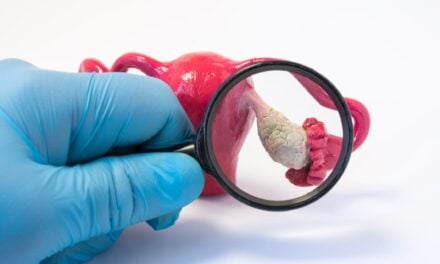
Engineers at Washington University in St. Louis have developed a microneedle patch that can be applied to the skin, capture a biomarker of interest and, thanks to its unprecedented sensitivity, allow clinicians to detect its presence.
The technology is low-cost and easy for clinicians or patients themselves to use, and it could potentially eliminate the need for a trip to the hospital just for a blood draw. In addition, the microneedle patches “are entirely pain free,” says Srikanth Singamaneni, PhD, the Lilyan and E. Lisle Hughes Professor in the Department of Mechanical Engineering & Material Sciences at Washington University. “They go about 400 microns deep into the dermal tissue. They don’t even touch sensory nerves.”
Finding a biomarker using these microneedle patches is similar to blood testing. But instead of using a solution to find and quantify the biomarker in blood, the microneedles directly capture it from the dermal interstitial fluid (ISF), the liquid that surrounds our cells in skin. Once the biomarkers have been captured, they’re detected in the same way—using fluorescence to indicate their presence and quantity.
ISF is a rich source of biomolecules, densely packed with everything from neurotransmitters to cellular waste. However, to analyze biomarkers in ISF, conventional method generally requires extraction of ISF from skin. This method is difficult and usually the amount of ISF that can be obtained is not sufficient for analysis. That has been a major hurdle for developing microneedle-based biosensing technology.
Another method involves direct capture of the biomarker in ISF without having to extract ISF. Like showing up to a packed concert and trying to make your way up front, the biomarker has to maneuver through a crowded, dynamic soup of ISF before reaching the microneedle in the skin tissue. Under such conditions, being able to capture enough of the biomarker to see using the traditional assay isn’t easy.
But the Washington University team has unique approach: “plasmonic-fluors,” an ultrabright fluorescence nanolabel. Compared with traditional fluorescent labels, when an assay was done on microneedle patch using plasmonic-fluor, the signal of target protein biomarkers shined about 1,400 times as bright and become detectable even when they are present at low concentrations.
“Previously, concentrations of a biomarker had to be on the order of a few micrograms per milliliter of fluid,” says Zheyu (Ryan) Wang, a graduate student in the Singamaneni lab. That’s far beyond the real-world physiological range. But using plasmonic-fluor, the research team was able to detect biomarkers on the order of picograms per milliliter. “That’s orders of magnitude more sensitive.”
These patches have a host of qualities that can make a real impact on medicine, patient care and research. For one, they would allow providers to monitor biomarkers over time, particularly important when it comes to understanding how immunity plays out in new diseases.
For example, researchers working on covid-19 vaccines need to know if people are producing the right antibodies and for how long. “Let’s put a patch on,” Singamaneni said, “and let’s see whether the person has antibodies against covid-19 and at what level.”
Another potential application would be during emergencies. “When someone complains of chest pain and they are being taken to the hospital in an ambulance, we’re hoping right then and there, the patch can be applied,” says Jingyi Luan, PhD, a recent graduate from the Singamaneni lab. Instead of having to get to the hospital and have blood drawn, EMTs could use a microneedle patch to test for troponin, the biomarker that indicates myocardial infarction.
Patches could also be used to monitor people with chronic health issues, eliminating the need for unnecessary trips to hospital.
Further research is necessary before the microneedle patches could be approved for clinical use. “We’ll have to determine clinical cutoffs,” that is, the range of biomarker in ISF that corresponds to a normal vs. abnormal level,” says Singamaneni. “We’ll have to determine what levels of biomarker are normal, what levels are pathological.”
Read more from Washington University in St. Louis.
Reference
- Wang Z, Luan J, Seth A, et al. Microneedle patch for the ultrasensitive quantification of protein biomarkers in interstitial fluid. Nat Biomedl Eng, 2021;5(1):64-76. doi: 10.1038/s41551-020-00672-y
Featured image: Engineers at the McKelvey School of Engineering at Washington University in St. Louis have developed a microneedle patch that can be applied to the skin, capture a biomarker of interest from interstitial fluid and, thanks to its unprecedented sensitivity, allow clinicians to detect its presence. Image Sisi Cao/Washington University in St. Louis.

