A study found that tracking tumor DNA in blood samples could identify signs of lung cancer recurrence months before they appear on scans, potentially helping physicians make earlier treatment decisions.
A blood-based liquid biopsy test can reliably detect microscopic traces of lung cancer in patients during the earliest clinical stages, identifying disease recurrence months before tumors appear on standard imaging scans, according to a study published in The Journal of Molecular Diagnostics.
The research demonstrates that tracking circulating tumor DNA (ctDNA) through serial blood draws accurately predicts relapse risk in patients treated for non–small-cell lung cancer (NSCLC). NSCLC accounts for the majority of lung cancer cases and remains the primary cause of cancer-related mortality. While patients with early-stage disease (stages I to II) often undergo curative surgical resection, disease recurrence rates range between 5% and 19% even in stage I, with periodic radiographic imaging serving as the current standard of care.
“As oncology shifts towards personalized risk-adapted care, liquid biopsies are emerging as a critical tool to bridge the gap between treatment completion and clinical recurrence,” says Matija Snuderl, MD, lead investigator from the Department of Pathology at NYU Langone Health and Grossman School of Medicine, and Laura and Isaac Perlmutter Cancer Center, New York, NY, in a release. “In our study, we monitored patients with lung cancer across treatment stages using serial blood draws to track circulating tumor DNA (ctDNA) levels. By analyzing tumor-specific signatures of ctDNA shed by tumor cells into the bloodstream, we evaluated how effectively liquid biopsy predicts relapse compared to standard imaging.”
Whole-Genome Sequencing and AI Analysis
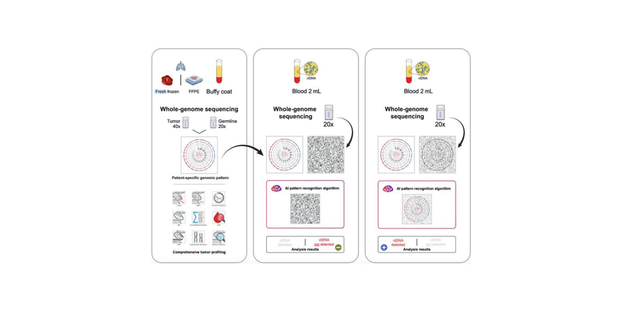
The monitoring assay uses whole-genome sequencing (WGS) technology developed by co-investigator Asaf Zviran, PhD, of C2i Genomics. In the laboratory workflow, tumor DNA is extracted from a formalin-fixed, paraffin-embedded pathology tissue sample, and normal germline DNA is isolated from the patient’s buffy coat. Both samples undergo WGS to establish a patient-specific mutational signature. During follow-up, ctDNA isolated from plasma undergoes WGS and is evaluated against the patient-derived signature using an artificial intelligence pattern recognition algorithm.
The study found that a positive test result for ctDNA served as a highly sensitive biomarker for minimal residual disease (MRD), identifying microscopic disease recurrence months ahead of physical manifestations on scans. Conversely, a clear or negative test result showed a strong correlation with sustained remission.
“As a surgeon, I follow my patients very closely, and I want to know as soon as possible if the cancer has come back. Unfortunately, even the earliest stage patients risk recurrence,” says Harvey I Pass, MD, co-lead investigator from the Department of Cardiothoracic Surgery at NYU Langone Health, in a release. “Our study’s findings highlight just how sensitive and specific liquid biopsy can be when integrated into routine post-treatment monitoring, even in the earliest stages of lung cancer. This is of critical importance for deciding on post-operative treatments.”
Clinical Implications for Post-Operative Management
Integrating sensitive MRD detection into routine laboratory testing provides multiple clinical advantages across the oncology care continuum. The liquid biopsy assay enables clinicians to stratify patients into high- and low-risk cohorts much earlier than conventional imaging protocols allow.
Detecting residual disease at microscopic levels allows oncologists to deliver targeted adjuvant therapies when tumor burden is minimal, improving long-term survival outcomes. Patients with sustained negative findings may be spared unnecessary adjuvant therapies, procedural toxicities, and the surveillance anxiety associated with waiting for routine imaging scans.
“Minimal residual disease testing via liquid biopsy represents a fundamental shift in post-operative care,” says Snuderl in a release. “It gives pathologists a powerful tool to diagnose disease before it appears on imaging and equips clinicians with actionable insights to personalize surveillance and intervention strategies for every individual patient.”
By identifying recurrence while disease burden remains low, the assay creates an opportunity for curative intervention before the cancer has a chance to spread, evolve and diversify, according to Asaf Zviran, PhD, of C2i Genomics.
Photo caption: Tumor DNA is extracted from a pathology sample, and normal DNA is extracted from the buffy coat. Both samples undergo WGS to generate the patient-specific mutational signature. At each follow-up, ctDNA extracted from plasma undergoes WGS and is analyzed using a patient-derived signature and an artificial intelligence (AI) pattern recognition algorithm. Formalin-fixed, paraffin-embedded: formalin fixed, paraffin embedded.
Photo credit: The Journal of Molecular Diagnostics / Snuderl et al