
Researchers at Washington University School of Medicine in St. Louis have developed a technique to detect minute amounts of a protein fragment linked to Alzheimer’s disease in the blood. The study shows that levels of p-tau-217 are elevated during the early stages of Alzheimer’s disease and could lead to a simple blood test capable of diagnosing the neurodegenerative disorder years before any symptoms begin to appear.1
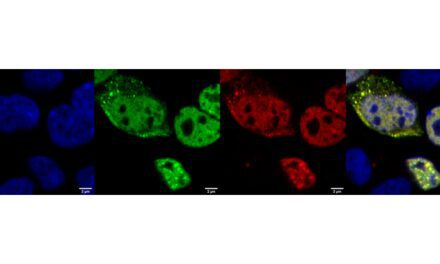
Alzheimer’s disease is characterized by the presence of plaques in the brain formed by a protein called amyloid-β, as well as aggregates of a protein called tau that form neurofibrillary tangles in the neurons of Alzheimer’s patients. Amyloid-β and tau start to change years before any cognitive symptoms, such as memory loss and confusion, become apparent, but previously the only way to detect them was with a positron emission tomography (PET) scan to visualize the brain or a spinal tap to measure changing levels of amyloid-β and tau in the cerebrospinal fluid. For many years, researchers have tried to develop blood tests that could detect Alzheimer’s disease before the onset of symptoms.
Randall Bateman, Nicolas Barthélemy, and colleagues at Washington University School of Medicine in St. Louis previously found that a modified fragment of tau, known as p-tau-217, accumulates in the cerebrospinal fluid of Alzheimer’s patients before the onset of cognitive symptoms, increases with disease progression, and can accurately predict the formation of amyloid plaques. The researchers suspected that p-tau-217 might also be present in the blood of Alzheimer’s patients, albeit at very low levels that would make it difficult to detect.
“We therefore wanted to quantify the levels of different tau proteins, especially p-tau-217, in the blood and compare them with amyloid pathology and onset of dementia to assess their potential as blood-based Alzheimer’s disease biomarkers,” says Bateman, who is the Charles F. and Joanne Knight Distinguished Professor of Neurology at Washington University School of Medicine. Barthélemy and colleagues in Bateman’s lab developed a mass spectrometry-based method to measure the amount of p-tau-217 and other tau fragments in as little as 4 ml of blood, even though such small samples may contain less than a trillionth of a gram of p-tau-217.
“To our knowledge, this is the lowest concentration ever measured by mass spectrometry for a protein marker in human blood plasma,” says Barthélemy, an assistant professor in Bateman’s laboratory. The researchers found that, similar to p-tau-217 levels in cerebrospinal fluid, p-tau-217 levels in the blood were extremely low in healthy volunteers but elevated in patients with amyloid plaques, even in those who had yet to develop cognitive symptoms. Measuring blood plasma levels of p-tau-217 was able to accurately predict the presence of amyloid plaques in PET scans, performing better than another tau fragment, p-tau-181, which was previously proposed as a biomarker for Alzheimer’s disease.
“Our findings support the idea that tau isoforms in the blood are potentially useful for detecting and diagnosing Alzheimer’s disease pathology,” Bateman says. “Moreover, our assay for measuring plasma tau levels could be used as a highly sensitive screening tool to identify tau changes associated with amyloid plaque formation in normal subjects, replacing costly PET imaging.”
Reference
- Barthélemy NR, Horie K, Sato C, Bateman RJ. Blood plasma phosphorylated-tau isoforms track CNS change in Alzheimer’s disease. J Exp Med. 2020;217(11):e20200861. doi:1084/jem.20200861


