
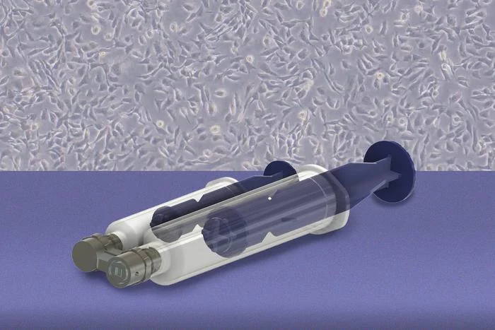
Using microfluidic shear stress, the system detaches living cells from excised tissue to support pathology workflows and downstream diagnostic testing.
Researchers at the Massachusetts Institute of Technology (MIT) and Johns Hopkins University have developed a handheld microfluidic device capable of gently collecting living cells from specific tissue locations to support cancer diagnostics, disease modeling, and personalized medicine development.
Described in a study published in the journal Device, the system enables targeted sampling of newly excised tissue without compromising surrounding tissue structures. The technique was developed to address persistent diagnostic hurdles in ovarian cancer, where high-grade serous cases frequently originate in the fallopian tubes as microscopic precursor lesions that are difficult to sample.
When ovarian cancer is identified in its earliest stages, five-year survival can exceed 90%. However, diagnoses that occur at stage 3 or stage 4 carry a five-year survival rate of less than half that figure.
“We wanted to collect living cells from specific regions of the fallopian tube while leaving the surrounding tissue intact,” says Kripa Varanasi, PhD, senior author of the study and the Maher A Elmasri professor of mechanical engineering at MIT, in a release. “Once we have these living cells, there are many things we can do with them. We can use them for diagnostics, grow them into organoids, and build living models of disease. Ultimately, this could allow us to test how an individual patient’s cells respond to different treatments and help us develop more personalized medicines.”
Microfluidic Shear Stress Mechanism
Standard pathology workflows typically place surgically excised tissue into chemical preservatives before slicing the specimen into thin sections for microscopic review. Although this process preserves cellular architecture, it renders cells nonviable, preventing laboratorians from cultivating them in culture or evaluating functional responses.
To address this constraint, the researchers designed a 3D-printed microfluidic device that interfaces directly with freshly removed tissue. The system uses one syringe to create a vacuum seal against the sample surface and a second syringe to direct fluid across a microfluidic channel. The localized fluid flow exerts a controlled shear stress parallel to the tissue surface, detaching living cells without requiring mechanical scraping with brushes or scalpels.
In comparative testing against standard cell-detachment approaches, cells gathered with the fluidic device maintained higher viability and grew in culture significantly more readily.
“This is exactly the kind of problem that benefits from bringing clinicians and engineers together,” says Rebecca Stone, MD, a gynecologic oncologist and the Stoddard and O’Neil professor in gynecologic oncology at Johns Hopkins University School of Medicine, in a release. “We understand the clinical need, while the MIT team brings a very different perspective from fluid mechanics and engineering. That combination allowed us to approach the problem in a new way.”
Applications Across Tumor Types and Pathology Workflows
The investigators validated the system on fresh human fallopian tube tissue. The team successfully isolated living cells and cultured them into organoid models before returning the remaining tissue intact for routine histopathology evaluation.
The device’s fluid shear forces can also be adjusted to match varying cellular adherence levels across different tissue types. For example, loosely adherent prostate cancer cells detach at approximately 1 pascal of stress, whereas bone cancer cells require up to 5 pascals of shear stress to detach.
“What is exciting about this technology is the ability to collect living cells from a specific area while preserving the tissue for pathology,” says Stone, in a release. “In the future, one could imagine integrating it into routine histopathology workflows, creating a powerful new way to study carcinogenesis and fundamental biology directly from human tissue.”
The researchers plan to focus initial clinical translation on newly excised surgical tissue specimens to streamline regulatory review. In future phases, the team aims to explore in vivo swabbing applications, which could permit direct sampling of internal patient tissues for earlier cancer detection.
The study was funded by the Break Through Cancer foundation, with additional co-authors including Domitille Avalle, Bert Vandereydt, Sean Parks, Huaiyao Peng, and Angela Belcher.
Photo caption: MIT researchers have developed a handheld device capable of gently collecting living cells from specific locations to test for ovarian and many other types of cancer.
Photo credit: Kripa Varanasi, et al