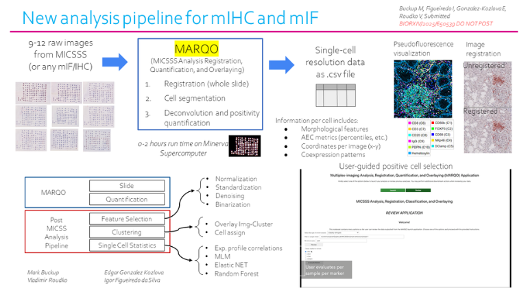
MARQO platform processes whole-slide immunohistochemistry and immunofluorescence images in minutes, potentially streamlining pathology workflows.
Researchers at the Icahn School of Medicine at Mount Sinai have developed MARQO, an artificial intelligence (AI)-powered computational tool designed to analyze cancer tissue slides with improved speed and accuracy compared to existing methods.
The platform, described in Nature Biomedical Engineering, processes immunohistochemistry (IHC) and immunofluorescence (IF) images to extract cellular and spatial information from tumor tissues. While currently designed for research applications, the tool’s compatibility with standard clinical staining methods suggests potential future applications in pathology laboratories.
“We designed MARQO to fill a major gap in the field: turning complex whole-slide images into usable, structured data quickly and consistently,” says Sacha Gnjatic, PhD, professor of immunology and immunotherapy at the Icahn School of Medicine and lead researcher, in a release. “By automating the heavy lifting, we let experts focus on interpretation and discovery.”
Addressing Current Analysis Limitations
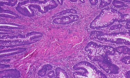
Traditional cancer tissue analysis requires pathologists to manually inspect stained tissue sections under microscopes to identify cell types and arrangements. This manual process is labor-intensive and typically limited to small sample areas, creating bottlenecks in workflow efficiency.
MARQO addresses these challenges through three key features: It processes entire slide images without requiring segmentation into smaller patches, completes analysis in minutes rather than hours using standard graphics processing units, and works across multiple common IHC and IF staining technologies to improve study reproducibility.
The platform automatically identifies likely positive cells and assigns coordinates and marker intensities, then provides this structured data to pathologists for final validation, maintaining human expertise in the diagnostic workflow.
Research Focus, Clinical Potential
The research team emphasizes that MARQO has not been validated for clinical diagnostics and remains a research tool. However, its design compatibility with standard clinical staining methods used in pathology labs suggests potential for future clinical applications.
The development team plans to enhance MARQO’s user interface, add advanced spatial and neighborhood analysis capabilities, and expand its use in high-performance computing environments to support large-scale projects involving millions of digitized tissue slides.
“This platform could accelerate biomarker discovery, improve how we predict which patients will benefit from specific treatments, and ultimately support the development of more precise cancer diagnostics,” says Gnjatic in a release.
The research received funding from the National Cancer Institute through U24, P01, and U01 grants, with additional seed funding from the Icahn School of Medicine at Mount Sinai.
Co-authors include Mark Buckup, now an MD/PhD student at University of California San Diego; Edgar Gonzalez-Kozlova, PhD, assistant professor of immunology and immunotherapy in the Gnjatic Lab; Igor Figueiredo, computer engineer; Pauline Hamon, PhD, now junior faculty in France; and Giorgio Ioannou, senior research assistant and lead on the multiplex IHC imaging platform.
Photo caption: MARQO delivers faster, fully integrated whole-slide image processing across multiple staining technologies
Photo credit: Mount Sinai


