
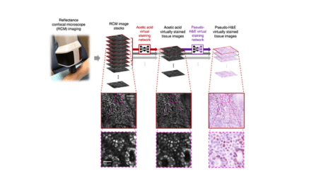
In an effort to push the level of performance for confocal imaging, a collaboration at the Marine Biological Laboratory (MBL) has invented a kitchen-sink confocal microscopy platform that successfully improves the its volumetric resolution by more than 10-fold while simultaneously reducing phototoxicity that has potential applications for imaging human tissue in histology and pathology labs.
This confocal microscopy platform borrows solutions from other high-powered imaging systems and adds a unifying thread of “Deep Learning” artificial intelligence algorithms.
The MBL’s report on the technology, called “Multiview Confocal Super-Resolution Microscopy,” was published in Nature.
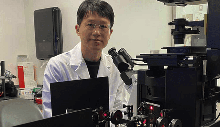
“Many labs have confocals, and if they can eke more performance out of them using these artificial intelligence algorithms, then they don’t have to invest in a whole new microscope. To me, that’s one of the best and most exciting reasons to adopt these AI methods,” says senior author and MBL Fellow Hari Shroff, PhD, of the National Institute of Biomedical Imaging and Bioengineering.
The new confocal platform uses three objective lenses, allowing one to image a wide variety of sample sizes, from nuclei and neurons in the C. elegans embryo to the whole adult worm. Multiple specimen views are rapidly captured, registered and fused to yield reconstructions with improved resolution over single-view confocal microscopy. The platform also introduces innovative scan heads for the three lenses, allowing line-scanning illumination to be easily added to the microscope base.
Moreover, the team added “super-resolution” capacity to the platform (enhanced resolution beyond the diffraction limit of light) by adapting techniques from structured illumination microscopy.
The team trained a deep learning computer model, or neural network, to distinguish between poorer-quality images with a low signal-to-noise ratio (SNR) and better images with a higher SNR. “Eventually the network could predict the higher SNR images, even given a fairly low SNR input,” says Shroff.
The team demonstrated the platform’s capabilities on more than 20 different fixed and live samples, targeting structures that ranged from less than 100 nanometers to a millimeter in size.
Yicong Wu, PhD, MS, first author on the paper, built the new confocal platform and deployed its Deep Learning approaches.
Featured Image: Mouse esophageal tissue slab (XY image), immunostained for tubulin (cyan) and actin (magenta), imaged in triple-view SIM mode. Photo: Yicong Wu and Xiaofei Han et al, Nature, 2021
