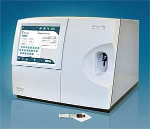

Nova Biomedical recently introduced the Nova pHOx Ultra analyzer, with 20 user-selectable tests.
Blood gas analysis and critical care medicine have been synonymous for many years. An important review published in the American Journal of Respiratory and Critical Care Medicine in 19981 makes that point. Its authors say that, “Critical care medicine is one of the newest and most rapidly growing medical specialties.” The paper elegantly retraces the development of knowledge regarding blood gas transport, the evolution of techniques to measure respiratory gases in whole blood, and how a polio epidemic in Copenhagen started the modern critical care movement and led to the a device that could measure pH in small volumes of blood directly. It should be noted that Radiometer Corp has changed the name of its educational Web site from bloodgas.org to acutecaretesting.org.
BACKGROUND: GAS EXCHANGE IN THE BLOOD AND LUNGS
The presence of both oxygen and carbon dioxide was documented in 1799 by Humphrey Davy. Nearly 40 years later, Heinrich Gustav Magnus found that there was more oxygen and less carbon dioxide in arterial blood than in venous blood. Jons Jacob Berzelius was able to split the red material found in blood into a protein called “globin” and a colored compound, which he named hematin, and showed that it took up oxygen. Felix Hoppe-Seyler renamed this compound “hemoglobin” in 1862. He found that oxygen formed a dissociable compound with hemoglobin that he called “oxyhemoglobin.” Carl Gustav von Hufner reported that 1.34 mL of oxygen combined with 1 g of crystalline hemoglobin, a number that has been essentially confirmed; the current published value being 1.39 mL.g. In 1917, Karl Hasselbach adapted Lawrence Henderson’s mass law for carbonic acid to a logarithmic form known as the Henderson-Hasselbach equation, which is fundamental in contemporary clinical acid-base analysis:pH =pK’ + log[HCO3– /SPCO2 ] where S = 0.0307
The first glass pH electrode was developed in 1925. Arnold Beckman perfected the first commercial pH meter in 1935. However, it was only used to measure acidity in citrus. A thermostated blood pH apparatus did not become commercially available until the mid-1950s. Today, prominent manufactures of blood gas analyzers include Siemens, Roche Diagnostics, Radiometer, Nova Biomedical, and Instrumentation Laboratory. Most manufacturers now produce small, portable stand-alone, easy-to-operate instruments designed for satellite or point-of-care operations. These instruments often combine systems for blood gases, and pH with ISEs and biosensors for electrolytes, glucose, urea nitrogen, creatinine, and hematocrit or hemoglobin. One recently introduced analyzer is the Nova pHOx Ultra, with 20 user-selectable tests including pH, PCO2, PO2, sO2%,Hct, Hemoglobin, Na+, K+, ionized calcium and ionized magnesium, Cl-, glucose, urea nitrogen, creatinine, lactate, CO-Oximetry, and total bilirubin. Some handheld devices such as the I-STAT use single-use disposable electrodes. Advances such as these have been made possible by the rapid evolution of microprocessors and the development of solid-state devices capable of detecting multiple analytes on a small surface.

CRAIG C. FOREBACK, PHD, is a senior lecturer, Department of Pathology & Laboratory Medicine, University of Wisconsin School of Medicine & Public Health. He wrote this article on behalf of Nova Biomedical.
WHY MEASURE BLOOD GASES?
Much has been written to answer the question, “Why measure blood gases?” Most clinical chemistry texts include information on clinical situations requiring the monitoring of blood gases and pH. Examples include Tietz: Fundamentals of Clinical Chemistry,2 and Clinical Chemistry Techniques, Principles, and Correlations.3 In a recent three-part introduction for the novice written by Chris Higgins4 (acutecaretesting.org, April, May, and June, 2012) part two addresses the question. Readers are directed to the Web site for detailed discussions. For the purposes of this discussion, the focus will be an explanation of the four classes of acid-base disturbances (five if you include mixed acid-base disorders), and the causes and consequences of these disturbances.
Given the wide range of medical conditions that may be associated with aberrations of acid-base balance, it is advisable to narrow the possible cause by classifying a patient’s acid base disturbance to one of four classes, which are: respiratory acidosis, respiratory alkalosis, metabolic acidosis, or metabolic alkalosis.
Respiratory acidosis is characterized by increased pCO2, which results in lowered pH and usually decreased pO2. This condition is invariably the result of hypoventilation due to chronic obstructive pulmonary disease, acute asthma, bronchopneumonia, or pulmonary edema. Disease or trauma to the chest wall and the musculature involved in the mechanics of respiration can result in hypoventilation. Certain drugs or a head injury can depress the respiratory center in the brain, which regulates the respiratory rate and can cause respiratory acidosis.
Respiratory alkalosis is characterized by decreased pCO2, which results in increased pH. It is always the result of hyperventilation that is often a feature of anxiety attacks. Excessive artificial ventilation has the same effect.
Metabolic acidosis is characterized by decreased [HCO3-], which results in decreased pH. Reduced bicarbonate is the defining feature of all cases. Increased production of metabolic acids, increased loss of bicarbonate from the body, or failure of the kidneys to regenerate bicarbonate can lead to a metabolic acidosis. In the critical care setting, metabolic acidosis is the most frequent acid-base disturbance leading to increased production of lactic acid. Any condition in which tissues are poorly oxygenated can cause increased production of lactate. In patients with diabetes, increased production of ketoacids is the cause of the metabolic acidosis that results from insulin deficiency or insulin inaction. In acute and chronic renal failure, reduced regeneration of bicarbonate by the kidneys and reduced urinary excretion of hydrogen ions is associated with metabolic acidosis.
Metabolic alkalosis is characterized by increased [HCO3-], which results in increased pH. This condition is commonly due to an abnormal loss of hydrogen ions from the body. Loss of gastric acid as a result of the vomiting of gastric contents or increased loss of hydrogen ions in urine due to excessive secretion of glucocorticoid or mineralocorticoid hormones can account for a metabolic alkalosis. Metabolic acidosis is common in patients with severe hypokalemia due to the movement of hydrogen ions from extracellular fluid in exchange for potassium ions. Conversely, alkalosis can lead to a redistribution of potassium causing a hypokalemia.
WHERE AND HOW SHOULD BLOOD GASES BE DONE?
Analysis of arterial blood gases can classify acid-base disorders into the four categories discussed. Many of the conditions that lead to these abnormalities are in patients needing critical care. Many medical facilities have chosen to set up blood gas instruments as satellite operations usually performed by respiratory therapists. In smaller hospitals, portable handheld devices may be used. Other hospitals prefer to perform blood gas analysis in a central laboratory facility, which can result in a longer turnaround time for results. Because of the critical nature of blood gas values in the intensive care setting, it would make sense to perform them at or near the ICU patient. In 2007, the National Academy of Clinical Biochemistry (NACB) released its final version of the practice guideline “Evidence-based Practice for Point-of Care Testing.”5 The document found persuasive evidence for improved patient outcomes with arterial blood gas testing in the ICU when used for the early detection and treatment of sepsis or shock.6
In another study of patients presenting to an urban emergency department and admitted to the ICU with either sepsis or shock, point-of-care testing was found to be effective. Using point-of-care methods to monitor blood gases and lactate reduced mortality when compared to patients receiving conventional treatment. The guideline also offered evidence that more rapid total turnaround time (TTAT) for blood gases, electrolytes, and magnesium leads to improved clinical outcomes in critical care patient settings, and that in point-of-care testing can be an effective way to reduce TTAT.
OTHER TEST PARAMETERS
The “what else” mentioned earlier refers to the comprehensive STAT menu that is available on blood gas/critical care analyzers. There are advantages to performing other analytes on such instruments. There is no need to draw another sample to obtain values for sodium, potassium, glucose, creatinine, lactate, ionized calcium, ionized magnesium, lactate, etc. Blood gas analyzers use direct potentiometry by ion-selective electrodes for the measurement of electrolytes. A primary advantage is that the technique is sensitive to molality and is therefore not affected by variations in the concentration of protein or lipids in the sample. Methods requiring sample dilution such as high-volume chemistry analyzers are affected by the presence of protein and lipids. In these methods, only the water phase of the sample is diluted, producing results lower than molality as a function of the concentration of protein and lipids in the sample. Thus, there is a risk for errors, such as a falsely low sodium (pseudohyponatremia), in cases of significantly elevated protein and lipid concentrations. Measurement of ions by direct potentiometry offers yet another unit of measurement known as activity (a), the concentration of free, unbound ion in solution.
Unlike methods sensitive to ion concentration, ISEs do not sense the presence of complexed or electrostatically “hindered” ions in the sample. This allows the measurement of ionized calcium and ionized magnesium. Physiologically, ionic activity is assumed to be more relevant than concentration when considering chemical equilibrium or biological processes.
The ability of the blood gas analyzer to determine other analytes such as urea nitrogen, creatinine, potassium, ionized calcium, and ionized magnesium is extremely important in light of the clinical conditions associated with acidosis and alkalosis. Given the increase in the incidence of diabetes and renal disease, the ability to determine creatinine in the renal failure patient and glucose in a patient in ketoacidosis is essential. Acidosis, alkalosis, and insulin therapy affect potassium values, necessitating the simultaneous monitoring of potassium along with blood gases.
Acidosis causes a transfer of intracellular potassium into the extracellular fluid, leading to hyperkalemia. Diabetic ketoacidosis and shock with tissue hypoxia can lead to hyperkalemia through extracellular redistribution. Potassium retention in acute renal disease and end-stage renal failure is yet another cause of increased plasma potassium levels. Alkalosis transfers potassium intracellular, leading to hypokalemis in plasma.
Studies have shown hypomagnesemia as illustrated by ionized magnesium levels is associated with higher morbidity and mortality rates. Soliman et al7 have concluded that development of ionized hypomagnesemia during an ICU stay is associated with a worse prognosis. A review by Shirey in the Journal of Anesthesia8 pointed out that 16% of heart surgery patients were hypomagnesemic. Patients receiving digoxin had preoperative total hypomagnesemia more frequently than patients not on digoxin. Ionized Mg was found to be a better predictor of disease than total magnesium in hypertension, AMI, head trauma, type 2 diabetes, stroke, ischemic heart disease, asthma, cyclosporine recipients, and in pregnancy. Manrique et al9 found that hypomagnesemia during cardiopulmonary bypass (CPB) in pediatric patients, as demonstrated by ionized magnesium methods, should be aggressively treated.
Brooks and Fry10 reported that approximately 24 hours after CPB plasma ionize, magnesium was significantly reduced, despite a small but significant increase in total magnesium. The Nova pHOx (pictured) ultra, StatProfile pHOx, and the Critical Care Xp are able to measure ionized magnesium. Measurement of ionized calcium is another important parameter along with blood gases whenever citrated blood products are administered (eg, liver transplants), and in patients with end-stage renal disease.
CONNECTIVITY
By connecting the ABL800 FLEX analyzer to Radiometer’s IT system, a user is able to control remotely placed analyzers throughout a hospital or even throughout a hospital network. Hospital-network integration is also possible through ASTM, HL7, and POCT1A standard communication protocols. Nova Biomedical’s Data Manager can consolidate multiple, networked pHOx Ultras into a comprehensive database for capture, archiving, and extensive data reporting, and also provides a single HIS/LIS interface for multiple pHOx Ultras throughout a network, eliminating separate interface costs. Remote review and remote control of multiple, network-connected analyzers can also be performed from an onboard Data Manager. Many of the handheld devices (Alere Epoc and Abbott i-stat1) have wireless capability.
THE FUTURE
As the acuity of patients seen in the hospital increases, point-of-care testing for blood gases and other critical care tests continues to expand. As a result, more and more blood gas/critical care analyzers are being placed at the point of care. For example, blood gas/critical care analyzers are playing a role in addressing the overcrowding crisis in the emergency department by providing improved turnaround time of urgent tests such as blood gases and chem.8 In many institutions, respiratory therapists are leading the way to improving patient care by providing more tests from a single sample using fewer resources and generating faster results.
Increased use of point-of-care testing also demands that devices be more automated. Blood gas analyzers will feature fully automated operation with analysis of selected test menus with just a push of a button. They will be able to perform an automated two-point calibration at preset intervals to assure that the instrument is ready for analysis at all times. Automated, onboard, true liquid quality control eliminates the steps involved in manually performing QC, thereby dramatically reducing labor costs.
Many analyzers now feature state-of-the-art biosensors that have been well characterized and proven to provide excellent accuracy throughout their analytical measurement range. Sophisticated, computerized self-monitoring of the entire analysis and calibration cycle assures error-free and accurate analyzer performance. Extensive automation of blood gas analyzers can eliminate variability due to operator technique. Another trend will be increased use of handheld devices that rely on a single-use cartridge. Driving this trend is the addition of more analytes on the disposable cartridge and the increasing demand for blood gas testing at the bedside and during air and ground transport of patients from remote locations to major medical centers.
Craig C. Foreback, PhD, is a contributing writer for CLP. For more information, contact Editor Judy O’Rourke at .
REFERENCES
- Severinghaus JW, Astrup P, Murray JF. Blood Gas analysis and critical care medicine. Am J Respir Crit Care Med. 1998;157:S114-S122.
- Burtis, Ashwood, Bruns, eds. Tietz Fundamentals of Clinical Chemistry, Sixth Edition. Philadelphia; Elsevier Inc; 2008.
- Bishop, Fody, Schoeff, eds. Clinical Chemistry Techniques, Principles, Correlations, Sixth Edition. Chapter 16.
- Higgins C. Why measure blood gases? A three part introduction for the novice. Available at: acutecaretesting.org. Accessed June 7, 2012.
- National Academy of Clinical Biochemistry. Laboratory Medicine Guidelines: Evidence based guidelines for point of care testing. Available at: www.aacc.org/SiteCollectionDocuments/NACB/LMPG/POCTLMPG.pdf. Accessed June 7, 2012.
- Rivers E, Nguyen B, Havstad S, et al. Early goal directed therapy collaborative group: Early goal directed therapy is the treatment of severe sepsis and septic shock. N Engl J Med. 2001;345:1368-1377.
- Soliman HM, Mercan, D, Lobo SS, Mélot C, Vincent JL. Development of ionized hypomagnesemia is associated with higher mortality rates. Crit Care Med. 2003;31(4):1082-1087.
- Shirey TL. Monitoring magnesium to guide magnesium therapy for heart surgery. J Anesth. 2004;18:118-128.
- Manrique AM, Arroyo M, Lin Y, et al. Magnesium supplementation during cardiopulmonary bypass to prevent junctional ectopic tachycardia after pediatric cardiac surgery: a randomized controlled study. J Thorac Surg. 2010;139(1):162-169.
- Brooks CIO, Fry CH. Ionized magnesium and calcium in plasma from healthy volunteers and patients undergoing cardiopulmonary bypass. Brit Heart J. 2003;69:404.
