The personal journey of co-author and physician Dana Mitchell details the challenges related to an accurate diagnosis of autoimmune encephalitis. Even as a medical professional, Mitchell faced significant roadblocks on her road to diagnosis and treatment.
By Dana Mitchell, MD, and Ilana Heckler, PhD
Autoimmune encephalitis (AE) remains an underappreciated neurological disorder that often poses a diagnostic and therapeutic challenge for patients and clinicians alike.
The Journey to an Accurate Autoimmune Encephalitis Diagnosis
My journey with autoimmune encephalitis began toward the end of my third year of medical school after I contracted a flu-like, viral infection while on one of my clinical rotations. Following resolution of the acute illness, I never felt as though I had fully recovered. I was experiencing fatigue and generalized weakness but attributed this to the rigors of medical school and my recent viral infection. Over the following weeks, I began to have daily morning headaches, which progressively worsened in frequency, intensity, and duration. Importantly, I did not suffer from headaches previously and could not remember a time when I needed to take medication to treat a headache.
As my headaches progressed, I began noticing other symptoms, including
- Left arm weakness
- Balance issues
- Blurry vision
- Pain with moving my right eye
- Heart palpitations
- Muscle twitches
- Parkinsonism
- Episodes of staring,
- Brain fog
At the time, I also had trouble organizing my thoughts and verbally communicating, making it extremely difficult for me to convey my concerns to providers. I can only describe the overall experience as though a “switch had been flipped,” a description I would later learn was common among autoimmune encephalitis patients and their family members.
At this point I already knew something was not right, but also worried that my symptoms were not “severe” enough for the cause to be identified with current diagnostic modalities. I also recognized that the breadth and fluctuating nature of my clinical symptoms, which I can best describe as neurological chaos, had the potential to be perceived as erratic and inconsistent, making my diagnostic journey even more challenging. My mother shared said that it seemed as though “every day brought symptoms that didn’t seem to be connected, but now it makes sense when you know that the main computer was malfunctioning. Her brain was irritated.”
Previously a skilled figure skater and honors medical student, in a matter of weeks, I found myself unable to subtract four from my own age or stand up in the shower without falling over. In addition, to the previously mentioned symptoms, a few key and/or infrequently described symptoms, stuck out to both me and my mother, further suggesting that something potentially serious was occurring:
- Characteristics of headaches: New onset, constant headache that progressively worsened in severity and frequency over a matter of days-weeks (pattern change or recent onset). It felt as though my brain was encased in shrink wrap that had been lit on fire. My headaches also became significantly more severe when laying down and bending over (positional headache) as well as with the Valsalva maneuver (coughing or straining). These findings, in addition to my neurological dysfunction (arm weakness, positive Romberg, cognitive deficits, etc.), are considered “red flags” and can suggest a secondary headache resulting from increased intercranial pressure caused by a mass (e.g. tumor, abscess), blood (e.g. stroke), or inflammation (e.g. meningitis or encephalitis), among other things1.
- Exaggerated and delayed acoustic startle response: While it is normal to experience some degree of response to sudden, loud noises, the onset of an excessive startle after infancy can be indicative of a serious neurological problem including, brainstem injury or neurodegeneration2-4. Importantly, exaggerated startle responses have been documented in patients with antibodies against glutamic acid decarboxylase (GAD) and the glycine receptor alpha-1 subunit (GlyRα1)4-6. Notably, over the course of my disease, I developed an exaggerated and delayed ASR, which progressively worsened over time. At first, I would experience more exaggerated muscle contraction, as though I became more “jumpy.” However, over the course of my disease, the intensity of my response increased, and the intensity of the stimulus needed to elicit this response decreased such that any sudden noise would evoke an abnormal startle response. In addition to prior reports regarding abnormal startle response secondary to anti-GlyRα1 and anti-GAD antibodies, previous studies have demonstrated that the acoustic startle response is modulated by key structures within the limbic system (the part of the brain responsible for regulating behavior, emotional responses and that is impacted in limbic encephalitis) including the amygdala, hippocampus, and anterior cingulate cortex7, 8. While the presence of a new, abnormal, reflexive response was enough for me to be concerned that something serious was going on, the specificity of this response to key areas of the brain and receptors known to be involved in antibody-mediated neurological disease (i.e. limbic encephalitis) heightened the suspicion for autoimmune encephalitis.
- Failure to perform the Stroop Test: The test is performed by presenting a picture (Figure 1) and asking the examinee to say the color of ink, rather than read the word. When the color of the ink does not match the written name of the color, the examinee takes longer and exhibits more errors than when the color name and color of ink are the same. Greater difficulty or an inability to perform this task has been reported in patients with dementia, neurodegenerative disease, and those with psychiatric disease.9 While the Stroop Test is not necessarily sensitive or specific for autoimmune encephalitis, my inability to complete this test, which I could previously complete without difficulty, provided some important pieces of information. Notably, successful performance on the Stroop test relies on proper functioning of a component of the limbic system, the anterior cingulate cortex, an area also known to be involved in regulating the acoustic startle reflex 7, 8, 10. Importantly, studies have shown that specific types of impairment on the Stroop test may be able to distinguish patients with purely psychiatric disorders from those with other causes of cerebral dysfunction.9

Nearly four weeks after I began noticing significant progression of my symptoms, I had an MRI study of my brain. At that time, the neuroradiologists reviewing the scans expressed concerns that I had bilateral hyperintensity (indicative of inflammation) of key components of my limbic system, including the anterior cingulate cortex, which is involved in processes and tasks that demonstrated impairment. While at the time no one knew precisely what this meant, the neuroradiologists expressed concerns that my scans were consistent with the early stages of encephalitis. Desperate to get better, I searched the literature and immediately discovered AE could be a very real cause of my symptoms. I shared this with my mother (a radiologist) who immediately recognized this to likely be my diagnosis.
I credit my successful diagnosis to my neuroradiologists who scoured the literature and consulted experts at centers with more experience to ensure proper evaluation of my scans. These providers, many of whom had never seen or treated this disease, but who were not dismissive of me or my mother when we discussed my clinical course and presented additional literature, were critical to achieving a full recovery.
Diagnostic Challenges in Autoimmune Encephalitis
Despite my relatively swift diagnosis within six months, the journey to obtain a correct diagnosis unfolded against a backdrop of several challenges, including:
- Bias in Medicine: The hysterical female
Potentially dangerous biases in healthcare can arise in the setting of AE due to several factors including a higher prevalence of the disease in young women, presentation of neuropsychiatric symptoms, a lack of knowledge about autoimmune encephalitis, and insufficient understanding of the underlying pathophysiology.
Even as a medical student with a mother who is a physician, I was not immune to bias, with my running diagnosis being “anxious, type A, female medical student” for the first six months of my journey. During an early hospital admission, a staff physician asked me, “Do you even want to be a doctor? Because it is really hard to be a physician and have babies.”
In practice, medical bias can result in critical tests not being ordered (e.g. MRI, PET scan, antibody panel, etc.) or results being overlooked. In fact, while I had an APE2 score ≥ 4 (sensitivity and specificity of 98% and 85% for an autoimmune etiology of epilepsy) and met all the requirements for either probable or definite autoimmune encephalitis over the course of my disease, a diagnosis of AE was not even considered by more than half of the physicians I initially saw.15, 16 Importantly, I exhibited signs of autonomic instability including significant fluctuations in blood pressure and heart rate, which had potentially dangerous implications, but were nonetheless frequently dismissed as anxiety. It was not until after my recovery when I stumbled across notes in my chart from New York, which highlighted my potential risk of “rapid decompensation secondary to autonomic instability,” that I realized the seriousness of this initial oversight and bias.
- Availability and Understanding of Clinical/Diagnostic Tests
In 2007, antibodies to anti-N-methyl-D-aspartate receptors (NMDAR) were first reported as a cause of autoimmune encephalitis.17 Since then, 21 additional antibody targets have been added to the Mayo Encephalitis Panel, with additional targets being discovered every year.
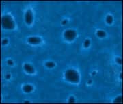
One of the diagnostic tests for AE utilizes a process called indirect immunofluorescence (Figure 2), which I use frequently in my own lab. In this process, the patient’s sample which contains antibodies, is incubated on a EUROIMMUN Autoimmune Encephalitis Mosaic 6 slide (AMPA-R, CASPR2-IgG, GABA-B-R, LGI1-IgG, NMDA-R) containing transfected and non-transfected HEK-293 cells. Alternately, the patient sample can be incubated on a slide containing frozen sections of mouse cerebellum, kidney, and gut tissues (Mayocliniclabs.com, TEST ID: ENC2 (CSF), ENS2 (Serum)). The sample is then incubated with another antibody, called a secondary antibody, which recognizes patient antibodies that have bound target antigens present on the tissue (e.g. NMDAR). This secondary antibody has a fluorescent label that can be visualized using a special type of microscope capable of exciting the fluorescent signal. As neuronal cell-surface antigens such as NMDAR are conformation dependent and fragile, solid phase detection methods such as Enzyme-Linked Immunosorbent Assay (ELISA) or immunoblot are not suitable.18 As such, the optimal method for the detection of autoantibodies against NMDAR is indirect immunofluorescence using recombinant cells which can be complemented by neuronal tissue sections and cultured primary neuronal cells.

While antibody testing is a helpful tool that can provide useful information capable of informing clinical decision-making, it is important for clinicians to understand how these tests are performed and the limitations of the current diagnostic modalities. Factors that can influence the detection of an antibody in a patient sample include:
- The strength and stability of the antibody-antigen interactions.
- The concentration of antibodies in a patient sample (which can be lower in early disease).
- The configuration of the antigen.
As such, it is possible for patients to have autoimmune encephalitis, but for current diagnostic tests to fail to identify a specific antibody. This is referred to as seronegative AE and prior work has suggested a set of clinical criteria to aid in the diagnosis of autoimmune encephalitis in patients who are found to be autoantibody negative on currently available tests16. In addition to the factors influencing antibody detection mentioned above, it is also possible that yet-to-be-identified pathogenic antibodies are the underlying cause of a patient’s disease, as would have been the case for NMDAR encephalitis patients prior to the identification of the anti-NMDAR antibody. Accordingly, an additional report by Souhel Najjar, MD, and colleagues, highlights key “red flag” clinical findings suggestive of an immune-mediated cause of neuropsychiatric disease, including AE19. Importantly, several recent studies suggest that clinical presentation and benefit of immunotherapy are not significantly different between patients with seropositive and seronegative autoimmune encephalitis20-23. As such, achieving optimal patient outcomes relies on providers appreciating the limitations of autoantibody testing and the importance of clinical symptoms in combination with radiographic findings to inform a final diagnosis.
MRI
MRI is often normal in autoimmune encephalitis.11 Notably, several recent publications have highlighted the superiority of [¹⁸F]fluorodeoxyglucose positron emission tomography ([¹⁸F]FDG-PET in the diagnosis of both seropositive and seronegative AE12-14, 23-27. Despite my MRIs being interpreted as abnormal both in the initial evaluation and subsequent evaluation at four independent institutions, skeptics persisted. Having come across early work by Probasco and Solnes et al.,12-14 I underwent evaluation by FDG-PET, which was determined to be grossly abnormal, becoming the most critical test in guiding my accurate diagnosis and subsequent treatment.
Blurring the Neurological-Psychiatric Divide
Autoimmune encephalitis is often characterized by the presence of a variable spectrum of, often fluctuating, neuropsychiatric symptoms, thus presenting a unique challenge for both patients and providers. Misdiagnosis of AE as a “purely psychiatric” disorder is not uncommon 28-34. I experienced this, with my earliest encounters resulting in the accumulation of psychiatric diagnoses including anxiety, depression, and functional neurological disorder (previously, conversion disorder) before I finally received the correct diagnosis in New York.
Clinically this is not unexpected, given the many similarities in presentation, but even more striking is the emerging research demonstrating a potential role for immune system dysfunction in a range of neuropsychiatric disorders, suggesting that beyond clinical findings, these disorders may share common pathophysiological underpinnings 35-37. Historically, the fields of psychiatry and neurology have existed as distinct specialties38. However, advances in our understanding of the brain and its function in health and disease over the last few decades has increasingly blurred the arbitrary line between these two disciplines38. Joseph B. Martin, MD, PhD, former dean of Harvard Medical School, has previously stated that “further progress in understanding brain diseases and behavior demands fuller collaboration and integration of these fields.” 38 It has been over two decades since Martin’s publication suggesting the need for closer integration of the neurological and psychiatric sciences38, yet barriers still remain and progress has been slow. In a more recent manuscript, Martin, Keshavan, and Price emphasize the persistence of “mind-body dualism” as well as “the complexities and uncertainties of psychiatry that have yet to translate into clinical practice,” as two key challenges yet to be overcome39.
Notably, a lack of understanding of the pathophysiological mechanisms underlying psychiatric symptoms, greatly impacts how medicine and society, view psychiatric versus neurological diseases. Fortunately, several key components can begin to aid in correcting and expanding our view:
- Addressing of bias in medicine (e.g. anxious type A female medical student) and reshaping how we think and talk about neuropsychiatric disease, beginning in medical school.
- Abolition of the concept of mind-body dualism.
- Awareness of knowledge gaps in the field as a whole and among individual providers. It is important to acknowledge the difference between, “I don’t know,” and “the field doesn’t know.”
- Improved and more expeditious integration and translation of pre-clinical research into clinical practice.
- Increased emphasis and funding of research in the fields of neurology and psychiatry.
About the Authors

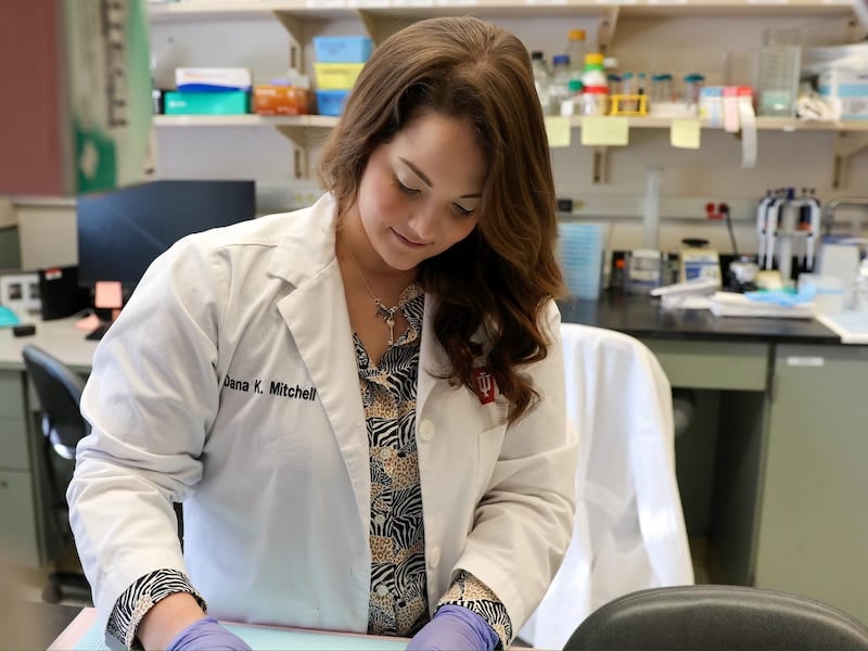
Dana Mitchell, MD, is currently an assistant research professor at the Indiana University School of Medicine in the Department of Pediatrics, where she serves as the chief of operations in the laboratory of Wade Clapp, MD, Chairman of the Department of Pediatrics and member of National Academy of Medicine.

Ilana Heckler, PhD, is a scientific affairs liaison at EUROIMMUN US (part of Revvity).
Featured Image: Since her successful autoimmune encephalitis diagnosis and treatment, co-author Dana Mitchell, MD, has continued her medical career in the Pediatrics Department at the Indiana University School of Medicine. Photo: Courtesy of Dana Mitchell
References
- Do, T. P.; Remmers, A.; Schytz, H. W.; Schankin, C.; Nelson, S. E.; Obermann, M.; Hansen, J. M.; Sinclair, A. J.; Gantenbein, A. R.; Schoonman, G. G., Red and orange flags for secondary headaches in clinical practice: SNNOOP10 list. Neurology 2019, 92 (3), 134-144.
- Saini, A. G.; Pandey, S., Hyperekplexia and other startle syndromes. J Neurol Sci 2020, 416, 117051.
- Henningsen, P.; Meinck, H. M., Specific phobia is a frequent non-motor feature in stiff man syndrome. J Neurol Neurosurg Psychiatry 2003, 74 (4), 462-5.
- Rauschenberger, V.; von Wardenburg, N.; Schaefer, N.; Ogino, K.; Hirata, H.; Lillesaar, C.; Kluck, C. J.; Meinck, H. M.; Borrmann, M.; Weishaupt, A.; Doppler, K.; Wickel, J.; Geis, C.; Sommer, C.; Villmann, C., Glycine Receptor Autoantibodies Impair Receptor Function and Induce Motor Dysfunction. Ann Neurol 2020, 88 (3), 544-561.
- Piquet, A. L.; Khan, M.; Warner, J. E. A.; Wicklund, M. P.; Bennett, J. L.; Leehey, M. A.; Seeberger, L.; Schreiner, T. L.; Paz Soldan, M. M.; Clardy, S. L., Novel clinical features of glycine receptor antibody syndrome: A series of 17 cases. Neurol Neuroimmunol Neuroinflamm 2019, 6 (5), e592.
- Bode, A.; Lynch, J. W., The impact of human hyperekplexia mutations on glycine receptor structure and function. Mol Brain 2014, 7, 2.
- Medford, N.; Critchley, H. D., Conjoint activity of anterior insular and anterior cingulate cortex: awareness and response. Brain Struct Funct 2010, 214 (5-6), 535-49.
- Lee, Y.; Davis, M., Role of the hippocampus, the bed nucleus of the stria terminalis, and the amygdala in the excitatory effect of corticotropin-releasing hormone on the acoustic startle reflex. J Neurosci 1997, 17 (16), 6434-46.
- Golden, C. J., Identification of brain disorders by the Stroop Color and Word Test. J Clin Psychol 1976, 32 (3), 654-8.
- Milham, M. P.; Banich, M. T.; Claus, E. D.; Cohen, N. J., Practice-related effects demonstrate complementary roles of anterior cingulate and prefrontal cortices in attentional control. Neuroimage 2003, 18 (2), 483-93.
- Kelley, B. P.; Patel, S. C.; Marin, H. L.; Corrigan, J. J.; Mitsias, P. D.; Griffith, B., Autoimmune Encephalitis: Pathophysiology and Imaging Review of an Overlooked Diagnosis. AJNR Am J Neuroradiol 2017, 38 (6), 1070-1078.
- Probasco, J. C.; Benavides, D. R.; Ciarallo, A.; Sanin, B. W.; Wabulya, A.; Bergey, G. K.; Kaplan, P. W., Electroencephalographic and fluorodeoxyglucose-positron emission tomography correlates in anti-N-methyl-d-aspartate receptor autoimmune encephalitis. Epilepsy Behav Case Rep 2014, 2, 174-8.
- Solnes, L. B.; Jones, K. M.; Rowe, S. P.; Pattanayak, P.; Nalluri, A.; Venkatesan, A.; Probasco, J. C.; Javadi, M. S., Diagnostic Value of (18)F-FDG PET/CT Versus MRI in the Setting of Antibody-Specific Autoimmune Encephalitis. J Nucl Med 2017, 58 (8), 1307-1313.
- Probasco, J. C.; Solnes, L.; Nalluri, A.; Cohen, J.; Jones, K. M.; Zan, E.; Javadi, M. S.; Venkatesan, A., Abnormal brain metabolism on FDG-PET/CT is a common early finding in autoimmune encephalitis. Neurol Neuroimmunol Neuroinflamm 2017, 4 (4), e352.
- Husari, K. S.; Dubey, D., Autoimmune Epilepsy. Neurotherapeutics 2019, 16 (3), 685-702.
- Graus, F.; Titulaer, M. J.; Balu, R.; Benseler, S.; Bien, C. G.; Cellucci, T.; Cortese, I.; Dale, R. C.; Gelfand, J. M.; Geschwind, M.; Glaser, C. A.; Honnorat, J.; Höftberger, R.; Iizuka, T.; Irani, S. R.; Lancaster, E.; Leypoldt, F.; Prüss, H.; Rae-Grant, A.; Reindl, M.; Rosenfeld, M. R.; Rostásy, K.; Saiz, A.; Venkatesan, A.; Vincent, A.; Wandinger, K. P.; Waters, P.; Dalmau, J., A clinical approach to diagnosis of autoimmune encephalitis. Lancet Neurol 2016, 15 (4), 391-404.
- Dalmau, J.; Bataller, L., [Limbic encephalitis: the new cell membrane antigens and a proposal of clinical-immunological classification with therapeutic implications]. Neurologia 2007, 22 (8), 526-37.
- van Coevorden-Hameete, M. H.; Titulaer, M. J.; Schreurs, M. W.; de Graaff, E.; Sillevis Smitt, P. A.; Hoogenraad, C. C., Detection and Characterization of Autoantibodies to Neuronal Cell-Surface Antigens in the Central Nervous System. Front Mol Neurosci 2016, 9, 37.
- Patel, A.; Meng, Y.; Najjar, A.; Lado, F.; Najjar, S., Autoimmune Encephalitis: A Physician’s Guide to the Clinical Spectrum Diagnosis and Management. Brain Sci 2022, 12 (9).
- Berger, B.; Hauck, S.; Runge, K.; Tebartz van Elst, L.; Rauer, S.; Endres, D., Therapy response in seronegative versus seropositive autoimmune encephalitis. Front Immunol 2023, 14, 1196110.
- Lee, S.; Kim, H. D.; Lee, J. S.; Kang, H. C.; Kim, S. H., Clinical Features and Treatment Outcomes of Seronegative Pediatric Autoimmune Encephalitis. J Clin Neurol 2021, 17 (2), 300-306.
- Lee, W. J.; Lee, H. S.; Kim, D. Y.; Lee, H. S.; Moon, J.; Park, K. I.; Lee, S. K.; Chu, K.; Lee, S. T., Seronegative autoimmune encephalitis: clinical characteristics and factors associated with outcomes. Brain 2022, 145 (10), 3509-3521.
- Roman, S. N.; Sadaghiani, M. S.; Diaz-Arias, L. A.; Le Marechal, M.; Venkatesan, A.; Solnes, L. B.; Probasco, J. C., Quantitative brain (18) F-FDG PET/CT analysis in seronegative autoimmune encephalitis. Ann Clin Transl Neurol 2024.
- Bordonne, M.; Chawki, M. B.; Doyen, M.; Kas, A.; Guedj, E.; Tyvaert, L.; Verger, A., Brain (18)F-FDG PET for the diagnosis of autoimmune encephalitis: a systematic review and a meta-analysis. Eur J Nucl Med Mol Imaging 2021, 48 (12), 3847-3858.
- Sadaghiani, M. S.; Roman, S.; Diaz-Arias, L. A.; Habis, R.; Venkatesan, A.; Probasco, J. C.; Solnes, L. B., Comparison of quantitative FDG-PET and MRI in anti-LGI1 autoimmune encephalitis. Neuroradiology 2023, 65 (8), 1225-1238.
- Kalra, S.; Tripathi, M.; Tripathi, M.; Sonar, R. S.; Pandey, A. K.; Jaleel, J.; Singh, R. K.; Kumar, P.; Damle, N. A.; Bal, C., Role of FDG PET/CT in definitive and presumed autoimmune encephalitis. Nucl Med Commun 2024, 45 (2), 121-127.
- Morbelli, S.; Djekidel, M.; Hesse, S.; Pagani, M.; Barthel, H., Role of (18)F-FDG-PET imaging in the diagnosis of autoimmune encephalitis. Lancet Neurol 2016, 15 (10), 1009-10.
- Xu, L.; Chen, Z., Anti-NMDA Receptor Encephalitis Misdiagnosed As Generalized Anxiety Disorder: A Case Report. Cureus 2021, 13 (12), e20529.
- Shimoyama, Y.; Umegaki, O.; Agui, T.; Kadono, N.; Minami, T., Anti-NMDA receptor encephalitis presenting as an acute psychotic episode misdiagnosed as dissociative disorder: a case report. JA Clin Rep 2016, 2 (1), 22.
- Ponte, A.; Brito, A.; Nóbrega, C.; Pinheiro, S.; Gama Marques, J., Catatonia in Anti-N-Methyl-D-Aspartate (NMDA) Receptor Encephalitis Misdiagnosed as Schizophrenia. Acta Med Port 2020, 33 (3), 208-211.
- Brummer, T.; Lotz, J.; Dresel, C.; Birklein, F., Anti-NMDA-receptor encephalitis and concurrent neuroborreliosis misdiagnosed for post-COVID-19-syndrome: a case report. Ther Adv Neurol Disord 2024, 17, 17562864231224108.
- Rainey, K.; Gholkar, B.; Cheesman, M., Anti-NMDA receptor encephalitis: an easily missed diagnosis in older patients. Age Ageing 2014, 43 (5), 725-6.
- Gurrera, R. J., Frequency and temporal sequence of clinical features in adults with anti-NMDA receptor encephalitis presenting with psychiatric symptoms. Psychol Med 2019, 49 (16), 2709-2716.
- Koksal, A.; Baybas, S.; Mutluay, B.; Altunkaynak, Y.; Keskek, A., A case of NMDAR encephalitis misdiagnosed as postpartum psychosis and neuroleptic malignant syndrome. Neurol Sci 2015, 36 (7), 1257-8.
- Khandaker, G. M.; Cousins, L.; Deakin, J.; Lennox, B. R.; Yolken, R.; Jones, P. B., Inflammation and immunity in schizophrenia: implications for pathophysiology and treatment. Lancet Psychiatry 2015, 2 (3), 258-270.
- Mondelli, V.; Blackman, G.; Kempton, M. J.; Pollak, T. A.; Iyegbe, C.; Valmaggia, L. R.; Amminger, P.; Barrantes-Vidal, N.; Bressan, R.; van der Gaag, M.; de Haan, L.; Krebs, M. O.; Nordentoft, M.; Ruhrmann, S.; Riecher-Rössler, A.; Rutten, B. P. F.; Sachs, G.; Koutsouleris, N.; McGuire, P., Serum immune markers and transition to psychosis in individuals at clinical high risk. Brain Behav Immun 2023, 110, 290-296.
- Drevets, W. C.; Wittenberg, G. M.; Bullmore, E. T.; Manji, H. K., Immune targets for therapeutic development in depression: towards precision medicine. Nat Rev Drug Discov 2022, 21 (3), 224-244.
- Martin, J. B., The integration of neurology, psychiatry, and neuroscience in the 21st century. Am J Psychiatry 2002, 159 (5), 695-704.
- Keshavan, M. S.; Price, B. H.; Martin, J. B., The Convergence of Neurology and Psychiatry: The Importance of Cross-Disciplinary Education. Jama 2020, 324 (6), 554-555.

