
A new research paper was published in Genes & Cancer, entitled, “Using quantitative immunohistochemistry in patients at high risk for hepatocellular cancer.”
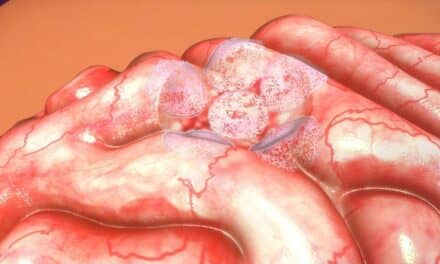
Hepatocellular cancer or HCC is the primary form of liver cancer and a major cause of cancer death worldwide. Early detection is key to effective treatment. Yet, early diagnosis is challenging, especially in patients with cirrhosis, who are at high risk of developing HCC.
Researchers Sobia Zaidi, Richard Amdur, Xiyan Xiang, Herbert Yu, Linda L. Wong, Shuyun Rao, Aiwu R. He, Karan Amin, Daewa Zaheer, Raj K. Narayan, Sanjaya K. Satapathy, Patricia S. Latham, Kirti Shetty, Chandan Guha, Nancy R. Gough, and Lopa Mishra from Northwell Health, The George Washington University, Georgetown University Medical Center, University of Hawaii, University of Hawaii Cancer Center, Zucker School of Medicine at Hofstra, North Shore University Hospital, University of Maryland, and Albert Einstein College of Medicine investigated changes in the transforming growth factor β (TGF-β) pathway for potential biomarkers that may differentiate hepatocellular cancer from cirrhosis.
The researchers determined that TGFBR2, not TGFBR1, was significantly reduced in hepatocellular cancer tissue compared with cirrhotic tissue. They developed an artificial intelligence (AI)-based process that correctly identified cirrhotic and HCC tissue and confirmed the significant reduction in TGFBR2 in HCC tissue compared with cirrhotic tissue.
In conclusion, the researchers proposed that a reduction in TGFBR2 abundance represents a useful biomarker for detecting hepatocellular cancer in the context of cirrhosis and that incorporating this biomarker into an AI-based automated imaging pipeline could reduce variability in diagnosing hepatocellular cancer from biopsy tissue.
The researchers noted that “[o]ur findings support further analyses to determine if this reduction is an early occurrence that can be used diagnostically in combination with other morphological characteristics or biomarkers to detect HCC at early stages in high-risk patients.”
Featured Image: AI-based analysis of TGFBR1 and TGFBR2 staining intensity in hepatocellular cancer and cirrhotic tissue. Image: Zaidi et al.

