LightDeck Diagnostics, Boulder, Colo, has been awarded a $5.65 million contract from the Biomedical Advanced Research and Development Authority (BARDA)—part of the Office of the Assistant Secretary for Preparedness and Response at the US Department of Health and Human Services (HHS)—to develop a rapid antigen test to detect SARS-CoV-2. These funds will enable LightDeck Diagnostics to develop a point-of-care antigen test that delivers results in less than 6 minutes.
“To stop the spread of COVID-19 and put an end to this pandemic, we need a public health strategy that includes national testing with widespread, and frequent, on-the-spot lab-quality testing,” says David Okrongly, chief science officer at LightDeck Diagnostics”
To detect infected individuals, rapid antigen tests represent an important adjunct to PCR testing, which can take days for results. The advantage of a rapid antigen test is that an infected individual can be detected and isolated in real time and contact tracing can be done right away.
The LightDeck Covid-19 Antigen Test leverages the highly sensitive and simple test procedure of waveguide technology with cost-effective manufacturing techniques to deliver an accurate, scalable test. The LightDeck antigen test will be a significantly faster alternative to the current PCR-based diagnostic tests and offer better sensitivity and specificity than the available lateral flow-based tests that require at least 15 minutes to deliver results.
“The LightDeck Covid-19 Antigen Test will build on the existing product platform,” says Chris Myatt, CEO and founder of LightDeck Diagnostics. “Every minute matters when we’re dealing with infectious diseases. We believe our technology can provide an optimal solution to the current gaps in covid-19 diagnostic testing on a national level.”
For more information, visit LightDeck Diagnostics.
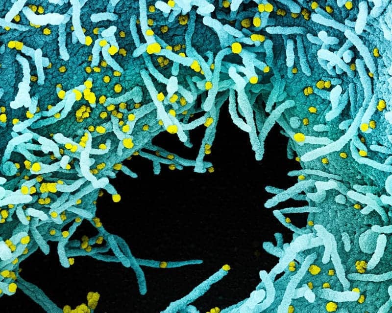
Featured image: Colorized scanning electron micrograph of a cell heavily infected with SARS-CoV-2 virus particles (yellow), isolated from a patient sample. The black area in the image is extracellular space between the cells. Image captured at the NIAID Integrated Research Facility (IRF) in Fort Detrick, Maryland. Credit: NIAID


