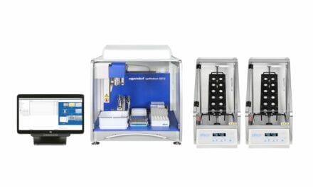
The CellaVision DM96 digital cell morphology system from Siemens, Malvern, Penn, automates traditional microscopy for high-volume hematology workflows in a hospital setting. Automatically locating cells on a stained peripheral blood or body fluid smear, the system preclassifies, stores, and presents the data for confirmation by a technologist to enhance speed and productivity. The system groups white blood cells into 18 discrete cell classes and precharacterizes red blood cells for polychromasia, hypochromasia, aniso-, micro-, and macrocytosis, and poikilocytosis. High-resolution images also detect cell inclusions. Users may compare different cell classes on one screen, while magnifying individual cells for closer analysis. A continuous slide feed raises throughput without increasing labor. For more information, visit Siemens.


