Applied Spectral Imaging (ASI), Carlsbad, Calif, a provider of biomedical imaging analysis solutions, has launched updated products designed to meet the growing demands of pathology and cytogenetic laboratories.
The product update includes a novel imaging methodology based on an all-new algorithm coupled with advanced hardware components to increase the scanning speed for fluorescence in situ hybridization (FISH) imaging. The new scanning methodology is designed to enhance various hematology and solid tumor imaging workflows.
By optimizing the traditionally error-prone process, ASI’s full-line of diagnostic products delivers laboratories with ultrafast sample scanning and highly efficient computer-assisted analysis for easier FISH validation, shorter turnaround times, and higher reporting confidence.

Limor Shiposh, Applied Spectral Imaging.
“We recognized that the rapid growth of FISH testing in pathology and increased test volumes have created a clear need for advanced digital workflows,” says Limor Shiposh, CEO of ASI. “I am excited to introduce our newly advanced imaging processor solution. It presents a paradigm shift in how FISH is performed, which is absolutely critical to patients.”
The new update has been incorporated into ASI’s brightfield, fluorescence, and spectral imaging and analysis solutions, including the company’s HiBand, HiFISH, Harmony, HiPath Pro, and PathFusion instruments.
PathFusion scanners for FISH, hematoxylin and eosinstained specimens, and immunohistochemistry imaging and analysis offer laboratories an all-in-one solution from scanning to reporting. In addition, the comprehensive pathology suite expedites more slide scanning volumes, improves diagnostic accuracy, and optimizes the overall workflow.
For further information, visit Applied Spectral Imaging.
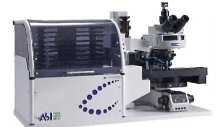
Featured image: Applied Spectral Imaging high-throughput tray loader and 99-slide high-throughput scanning system, which can automate the handling of more than 2,000 slides per week.
