By Louise Lazear
 Diagnosing lupus may become easier with the development of new testing methods
Autoimmune disease encompasses a plethora of disorders in which the body produces an immune response against its own tissues. Because these disorders often are associated with symptoms similar to those of other etiologies, criteria were developed in the 1970’s to assist the medical community in classifying diseases as autoimmune in nature. These include: findings in the blood and tissue affected by the disease that are consistent with inflammatory or immunological processes; the existence of autoantibodies to specific proteins normally found in tissue; and the presence of immune response cells (i.e. T lymphocytes) in affected tissues. While these autoimmune disorders are estimated to cost $86 billion per year, autoimmunity as a major disease category has yet to be accepted by the medical community. Some feel this stems from the fact that diagnosis is often complicated and lengthy, as autoimmunity is often overlooked as an underlying cause of symptoms. However, new assays for autoimmunity and immunosuppressive therapies for some diseases may help to change the way clinicians currently address these disorders.
Autoimmune diseases fall into two main categories: systemic or localized. Examples of systemic types include rheumatoid arthritis, lupus, scleroderma, and Sjogren’s syndrome. Localized diseases include Crohn’s disease, multiple sclerosis, Graves’ disease, and autoimmune hepatitis. While the cause of autoimmune disease remains unknown, there appears to be both a genetic predisposition and environmental trigger for virtually all the disorders. According to the American Autoimmune Related Diseases Association (AARDA), an estimated 50 million people in the U.S. suffer from about 80 classified autoimmune diseases. While many of these disorders are not life threatening, a recent study concluded that autoimmune diseases are the fifth leading cause of death by disease in women aged 15 to 44. Autoimmunity has been shown to be more prevalent in women than men, and female to male ratios of various diseases are compelling: 50 to one for hypothyroiditis, and nine to one for systemic lupus erythematosus (SLE). However, people of African, Asian and American Indian descent appear to be afflicted with autoimmune disease even more frequently than Caucasian woman.
Lupus, the hallmark immune complex disease, is a chronic inflammatory state that affects the skin, joints, blood and kidneys. Clinically, lupus runs the gamut from a mild butterfly rash on the nose and cheeks to a life-threatening disease state. It is estimated that between 500,000 and one million people in the U.S. have been diagnosed with this disease. Lupus falls into three general categories: discoid (cutaneous), systemic, and drug-induced. The most severe form is systemic lupus erythematosus (SLE), which can affect almost any organ in the body. The most common symptoms of SLE, which are often cyclic in nature, include arthralgia, fever, arthritis, fatigue and skin rashes. While causes of SLE remain unknown, in 1997 researchers isolated a gene on chromosome 1 that may predispose people to SLE. However, statistics indicate that no more than 5 percent of people with parents diagnosed with SLE will develop the disease. Some scientists have determined that environmental factors such as infection, ultra-violet light and hormones can also trigger the disease.
|
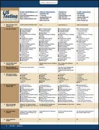
Testing Algorithm Using IFA As Screening Method
|

There is no definitive diagnostic marker for SLE. Because symptoms vary between patients as well as during the course of the disease, diagnosis and disease management is dependent upon the patient’s entire clinical presentation. Diagnosis involves a complex algorithm based on patient history, physical examination and various diagnostic assessments for abnormalities in the blood and kidneys as well as measurements of autoimmunity. Blood cell anomalies often occur in patients with SLE, and diagnosis often includes testing for anemia, leukopenia, and thromobocytopenia. Lupus commonly affects the kidneys, and renal function is evaluated via creatine clearance measurement to determine glomerular filtration rate. Urinalysis for proteinuria and the presence of RBCs, WBCs and cellular fragments can also indicate renal involvement. Serum creatinine concentration, another indicator of renal impairment, and kidney biopsy may also be used at various stages in a work-up for SLE. Therapies for SLE include topical applications of anti-inflammatory agents, as well as powerful steroids and new immunomodulating drugs that may reduce the need for steroid administration and associated side effects.
Measurements of autoimmunity by testing for the presence of certain autoantibodies can diagnose SLE and help differentiate SLE from other autoimmune disorders. Typically, physicians will order an antinuclear antibody (ANA) screen to rule-out SLE, as a positive ANA result is found in close to 100 percent of patients with active disease. However, positive results are also found in 95 percent of patients with mixed connective tissue disease, and false positives can occur as a result of chronic infectious disease and exposure to certain drugs. Negative ANA results typically do not require further autoimmune work-up.
Sensitivity and specificity of ANA measurements are related to the type of assay employed. Many laboratories use indirect immunoflourescent (IFA) with a human epithelial (HEp-2) cell line as substrate. Samples are incubated with the substrate, washed, then incubated with flourescein-labeled conjugate. When examined under fluorescent microscopy, positive samples exhibit green fluorescence in the cytoplasm or nuclei which demonstrate bound antibody. Specific stain patterns have been associated with some autoimmune disorders. Homogenous or rim-staining patterns and high titers are indicative of SLE, while a centromere pattern is often associated with Crest syndrome. Speckled and nucleolar patterns are associated with Sjogren’s syndrome and diffuse scleroderma respectively, but may also be indicative of SLE in some cases. IFA, while very sensitive for SLE, is also subjective and labor intensive. With the advent of new ELISA-based ANA screens, many large volume laboratories reflex positive specimens to IFA to determine stain pattern and titer.
Positive ANA screens may then be followed with tests specific for ANAs that are known to have high association with SLE, including anti-dsDNA (double-stranded), anti-SSA (Ro), anti-SSB (La) and anti-Sm (Smith). Anti-dsDNA is highly specific for SLE, occurring in 60 to 80 percent of patients with active disease. Anti-dsDNA, which is not found in patients with other autoimmune disease, correlates with lupus nephritis. Measurement of anti-dsDNA can be used to monitor disease activity and therapy response, and can also be of prognostic value.
| Survey Shows Lupus Is Difficult to Diagnose
A survey conducted by the Lupus Foundation of America suggests that more than half of the people afflicted with lupus suffer at least four years, and see three or more doctors before obtaining a correct diagnosis, reinforcing the need for greater awareness of lupus symptoms among patients and doctors alike. The survey of more than 1,000 LFA members also revealed that two of three lupus patients experience complete or partial loss of their income because they are unable to work, and that one in three are permanently or temporarily disabled by the disease. One of three patients responding to the LFA survey reported they had another autoimmune disease in addition to lupus, and almost half had another family member afflicted with an autoimmune disease. Nearly half of the survey participants (49%) received their diagnosis of lupus after being examined by a rheumatologist, a medical specialist who treats diseases of the connective tissue. Four of ten lupus patients are being treated by three or more doctors, and take six or more medications to treat symptoms of the disease. Most reported they are coping well with lupus (78%), and that other family members are understanding and supportive (72%). People with lupus named other family members (84%) and friends (72%) as their primary support network. The survey participants cited pain (65%), lifestyle changes (61%), and emotional problems associated with lupus (50%) as the most difficult factors for coping with lupus. For more information visit the Lupus Foundation of America website at www.lupus.org. |
Assays for anti-chromatin and anti-histone in serum can also be beneficial in the diagnosis of SLE and drug-induced lupus (DIL). Chromatin, the building block of chromosomes, is a DNA/histone complex found in the cell nucleus. Researchers have found that between 50 to 90 percent of SLE patients are positive for anti-chromatin antibodies, and that the presence of anti-chromatin antibodies may be more sensitive and specific for DIL than the presence of anti-histone. “Testing for chromatin is far more sensitive for lupus than testing for anti-DNA antibodies,” said Brys Myers, vice president of marketing at Inova Diagnostics, Inc. “Chromatins were the first autoantibodies ever discovered. They are responsible for the LE cell phenomena. Assays for chromatin have replaced the LE cell test, which was very labor intensive, yet extremely specific. People will still continue to test for anti-DNA, however, particularly because of its historical value,” he added.
SSA, SSB, Sm, SmRNP, Scl-70, and Jo-1 are nonhistone proteins that are typically referred to as extractable nuclear antigens, or ENAs. Several ENA profiles are commercially available and feature various combinations of these antigens to test for the presence of autoantibodies. Again, positive ENA profiles may then reflex to specific antigen assays to differentiate between disorders. Although only 30 percent of patients with SLE test positive for Anti-Sm, it is rarely associated with other autoimmune diseases and is therefore highly specific for SLE. Anti-SSA and anti-SSB are usually present together, and can be useful in differentiating SLE and Sjogren’s syndrome from other disease. Total hemolytic complement (CH50) can be used as a general screen for abnormalities relating to the entire complement system, and specific measurements of C3 and C4 complement can aid in determining renal involvement, disease progression, and response to therapy.
Antiphospholipid antibodies (APLs), which are present in 30 to 40 percent of SLE patients, are autoantibodies that react with most negatively charged phospholipids including prothrombin and beta2-glycoprotein1. A positive APL along with thrombosis, recurrent fetal deaths, or thrombocytopenia, leads to a diagnosis of APL syndrome. Various assays have been developed to detect APLs, including an ELISA assay for anticardiolipin antibody (ACA). “The anti-cardiolipin antibody (ACA) was first detected in a patient with lupus,” said Luis Lopez, M.D. and CEO of Corgenix, Inc. “The cardiolipin test is viewed by many in the field as a screening test, similar to the ANA screen. If the test is positive, you would continue to perform tests for anti-beta2 glycoprotein 1 and anti-prothrombin,” he said. Positive secondary tests can lead to a diagnosis of antiphospholipid syndrome secondary to lupus if the patient also presents with thrombosis. According to Lopez, presence of APLs can also indicate an increased risk for thrombosis in patients with SLE.
Because some APLs also inhibit activated partial thromboplastin time and other coagulation studies, they have come to be known as “lupus anticoagulants” despite the fact that clinical manifestations include thrombosis and other components of the APL syndrome. Most of these autoantibodies are directed against prothrombin or beta2-glycoprotein 1. Positive lupus anticoagulant tests can also indicate an increased risk of thrombosis for SLE patients.
Even to those well versed in autoimmune disease, it is understandable why diagnosis is often lengthy and complex. A new multi-plexing platform that allows for quantitative as well as qualitative measurement of up to 100 analytes per sample is the framework for two recently FDA 510(k) cleared autoimmune assays developed by Inova Diagnostics and Zeus Scientific, with others soon to follow. These assays use the Luminex xMAP system, an open-platform technology that employs microspheres labeled with specific antigens combined together in the same well. Unlike ELISA, each anti-body/antigen microsphere is measured individually, so quantitative as well as qualitative determinations are available. Inova’s ENA assay provides measurement of antibodies to Sm, RNP, SSA and SSB. According to Inova’s Myers, the company has additional assays under development, including more specific panels for liver and vasculitis. Although excited about the potential of this new platform, Myers acknowledged that the cost of the technology could limit its use to large volume settings. “There are significant upfront costs, including the cost of the system and the price of the beads (reagent). You have to run a lot of samples to make it cost effective. And until the bead price comes down, certain assays, although they work quite well, may not be feasible.”
The Zeus AtheNA Multi-Lyte ANA test system, also based on the Luminex platform, provides semi-quantitative detection of antibody to SSA, SSB, Sm, RNP, Scl-70, Jo-1, centromere B and histone, as well as qualitative detection of ANAs and quantitative detection of anti-dsDNA. “This is a tremendous savings for the hospital in terms of labor, and clinically you get so much more information up front,” said Jim Meenan, product manager at Wampole Laboratories which is distributing the system. “We are estimating up to a 95 percent savings in labor in the laboratory. This could revolutionize autoimmune testing from that perspective,” he said.
Other companies including Bio-Rad Laboratories and Immunoconcepts have entered into strategic partnerships with Luminex to develop the xMAP technology and bring additional products to market. “The technology is being commercialized in many different areas”, said Ralph McDade, COO and vice president of R&D at Luminex Corporation. “If there has ever been a paradigm shift in the lab, this is it”, he added. Wampole’s Meenan agrees. “I fully suspect over the next several years we will see much of diagnostic testing move to multi-plexing platforms. Allergy testing, for example, would seem like a natural fit,” he added.
Like any new medical technology, issues of cost and reimbursement will impact eventual utilization. But for now, multiplexed ANA and ENA platforms could help reduce the complicated and often lengthy process leading to diagnosis of SLE and other autoimmune diseases.