
A quick and easy test to detect cancer from blood or biopsy tissue could eventually result in a new approach to patient diagnosis. The test has been developed by University of Queensland researchers Abu Sina, PhD, Laura Carrascosa, PhD, and Matt Trau, PhD, who have discovered a unique DNA nanostructure that appears to be common to all cancers.1
Cancer is an extremely complicated and variable disease and different types of cancer have different signatures. Consequently, it has previously proven difficult to identify a simple signature that is distinct from healthy cells yet common to all cancers. The newly discovered nanostructure appears to fill the bill.
“This unique nano-scaled DNA signature appeared in every type of breast cancer we examined, and in other forms of cancer, including prostate, colorectal, and lymphoma,” says Sina. “The levels and patterns of tiny molecules called methyl groups that decorate DNA are altered dramatically by cancer. These methyl groups are key for cells to control which genes are turned on and off.”
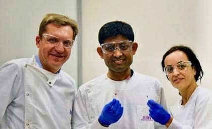
University of Queensland researchers (from left): Matt Trau, PhD; Abu Sina, PhD; and Laura Carrascosa, PhD.
The researchers took a holistic approach and developed a tool that could look at the nanoscale pattern changes at the whole-genome level within minutes. “In healthy cells, these methyl groups are spread out across the genome,” says Carrascosa. “But the genomes of cancer cells are essentially barren except for intense clusters of methyl groups at very specific locations.”
Cancer cells release DNA fragments into blood plasma when they die. The team discovered that intense clusters of methyl groups placed in a solution caused these cancer DNA fragments to fold into unique three-dimensional nanostructures that could easily be separated by sticking to solid surfaces such as gold.
“We designed a simple test using gold nanoparticles that instantly change color to determine if the 3D nanostructures of cancer DNA are present,” says Trau. “So we were very excited about an easy way of catching these circulating free cancer DNA signatures in blood.
“Discovering that cancerous DNA molecules formed entirely different 3D nanostructures from normal circulating DNA was a breakthrough that has enabled an entirely new approach to detect cancer noninvasively in any tissue type, including blood,” adds Trau. “This led to the creation of inexpensive and portable detection devices that could eventually be used as a diagnostic tool, possibly with a mobile phone.”
The new technology has proved to be up to 90% accurate in tests involving 200 human cancer samples and normal DNA.
“We certainly don’t know yet whether it’s the holy grail for all cancer diagnostics, but it looks really interesting as an incredibly simple universal marker of cancer, and as an accessible and inexpensive technology that doesn’t require complicated lab-based equipment like DNA sequencing,” says Trau.
The researchers are working with UniQuest, the University of Queensland’s commercialization company, to further develop the technology and license with a commercial partner.
Reference
- Sina AAI, Carrascosa LG, Liang Z, et al. Epigenetically reprogrammed methylation landscape drives the DNA self-assembly and serves as a universal cancer biomarker. Nature Communications. 2018;9:4915; doi: 10.1038/s41467-018-07214-w.
Featured image: Gold substrate with cancer DNA represented by spikes. Image courtesy University of Queensland.


