
Olympus Corp, Waltham, Mass, has recently described the findings of a research program that has the potential to help streamline the workload of clinical pathologists through the use of digital imaging and artificial intelligence systems.
The research program, “A New Approach to Develop Computer-Aided Diagnosis Using Artificial Intelligence for Gastric Biopsy Specimens,” was conducted jointly with Kiyomi Taniyama, MD, PhD, president of Japan’s Kure Medical Center and Chugoku Cancer Center. Initiated in 2017, the research program coupled Taniyama’s expertise in diagnostic pathology of the gastric system and digital pathology with Olympus’ imaging system technology and proficiency in artificial intelligence development.

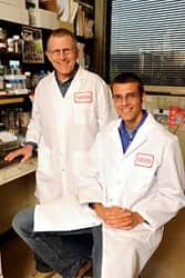
Kiyomi Taniyama, MD, PhD, Kure Medical Center and Chugoku Cancer Center.
Making use of the company’s proprietary deep-learning technology, Olympus has been working to develop a computer-aided diagnostic solution using artificial intelligence for diagnostic pathology.
For the research program, Olympus applied its deep-learning technology to a review of gastric biopsy specimens collected at the Kure Medical Center and Chugoku Cancer Center between 2015 and 2018. The outcome was the creation of a multiresolutional convolutional neural network (CNN), a network structure that is widely used in deep-learning technology for image analysis.
The deep-learning technology’s unique CNN was designed to analyze the features shown in digital images of pathology specimens. Such a structure effectively realizes the learning features of input data.

Table 1. When the threshold of the computer-aided diagnostic system was set to assess all 297 ADC samples as positive (100% sensitivity), 225 out of the 489 NADC samples were assessed as negative. This represents 100% sensitivity (297 out of 297) and 46% specificity (225 out of 489).
The research program was executed in two stages. In the learning stage, the artificial intelligence system used digital pathology images and associated information to ‘learn’ the CNN model. In the prediction stage, the artificial intelligence system applied its learnings about the CNN model to classify images of tissue specimens as either adenocarcinoma (ADC) or non-adenocarcinoma (NADC).
The artificial intelligence algorithm was developed using 368 whole-slide pathology images for learning, and 786 sample images (297 ADC; 489 NADC) to fine-tune the classification thresholds (Table 1). In whole-slide imaging, pathology specimens on glass slides are digitally imaged in their entirety and displayed for view on a monitor.
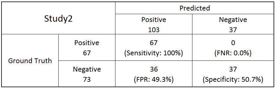
Table 2. A final evaluation of the computer-aided diagnostic system was made using 140 previously unseen sample images (67 ADC; 73 NADC). All 67 of the ADC samples were assessed as positive; 37 out of the 73 NADC samples were assessed as negative. These results represent 100% sensitivity (67 out of 67) and 50.7% specificity (37 out of 73).
The classification threshold established for the detection of all ADC samples achieved 46% specificity with NADC samples. In a final evaluation performed with 140 new cases (67 ADC; 73 NADC), the same algorithm and classification threshold achieved 100% sensitivity and 50.7% specificity (Table 2).
Computer-aided diagnostic software using artificial intelligence to achieve a low rate of false-negative results—that is, positive results incorrectly assessed as negative—can help pathologists detect positive samples.
The software developed by Olympus has the potential to eliminate duplication of effort in the workload of pathologists and further improve the accuracy of pathology diagnosis of gastric biopsies by screening negative samples and helping prevent positive samples from being overlooked.
For further information, visit Olympus.
Featured image: Gastric adenocarcinoma. Microphotograph by Viachaslau Bondarau courtesy Dreamstime.


