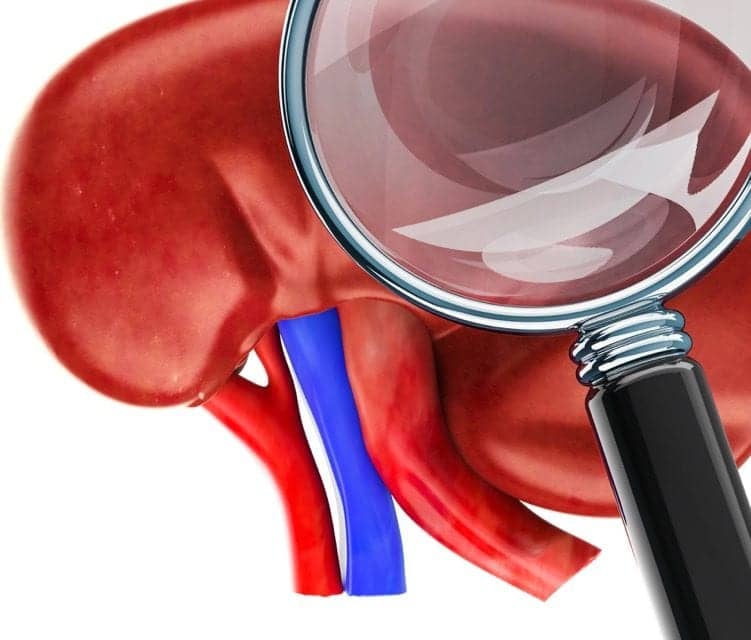
A research group from the Medical University of Vienna has successfully described the histological features of urinary obstruction in humans the first time. Obtained from kidney transplant patients, the findings may make it possible in the future to identify potentially dangerous complications following a kidney transplant at an earlier stage than currently, and thus provide prompt treatment.1
Obstructive urological complications can occur following a kidney transplant and can lead to urinary obstruction and subsequently to allograft failure. These complications include narrowing of the ureter, a leak between ureter and urinary bladder, or a hematoma that compresses the allograft or the efferent urinary tract. Although such complications can normally be detected by ultrasound, in the first few months following a kidney transplant this test is not always sufficiently diagnostically conclusive for detecting urinary obstructions. Where there is no clear diagnosis for restricted renal function following a transplant, a pathohistological workup of a kidney biopsy is therefore essential to establish the cause so that appropriate treatment can be started.
“Histology results are one of the most important diagnostic tools in clarifying the cause of restricted renal function,” says lead author Marija Bojic, MD, from the department of internal medicine III at the Medical University of Vienna. “Unfortunately, the existing literature on the subject inadequately describes the histological criteria that indicate a urinary obstruction.”
The present study enabled the study team, led by Zeljko Kikic, MD, also of the department of internal medicine III at the Medical University of Vienna, to describe a specific histological phenotype that is associated with just such obstructive urological complications.
The study involved the examination of biopsies from 976 kidney transplant patients. In particular, the researchers were looking for the presence of ‘tubular ectasia,’ or distension of the renal tubules. “These changes were observed in earlier animal models where a urinary obstruction was stimulated,” says Kikic. “We therefore wanted to investigate whether this also occurs in humans.”
In addition, further changes in the renal tubules were analyzed and the results correlated with the existence of proven obstructive urological complications. The Vienna researchers have now mapped out and described exact features that indicate a urinary obstruction.
“These results can be used for the earlier detection of occult urinary obstructions so that patients can receive the necessary treatment more quickly,” says Bojic. The results have been published and presented at the international conference of the Banff Foundation, Pittsburgh, which is playing a major role in drafting international guidelines on allograft rejection.
For more information visit Medical University of Vienna.
Reference
- Bojic M, Regele H, Herkner H, et al. Tubular ectasia in renal allograft biopsy: associations with occult obstructive urological complications. Transplantation. Epub ahead of print, March 4, 2019; doi: 10.1097/tp.0000000000002699.
Featured image:
Kidney research. Image © Alexlmx courtesy Dreamstime (ID 132282909).


