Summary: The descSPIM is an innovative, low-cost light sheet fluorescence microscopy (LSFM) system developed by researchers at Juntendo University for 3D imaging of cleared tissue samples, significantly enhancing accessibility for biomedical research.
Takeaways:
- Accessibility and Cost: descSPIM reduces the cost of LSFM systems to between $20,000 and $50,000, making high-quality 3D imaging more accessible.
- Validation and Application: The system successfully imaged various tissues, including brain and tumor samples, demonstrating its potential for detailed biological research.
- Open-Source Design: The open-source nature of descSPIM fosters collaborative development and widespread adoption in the scientific community.
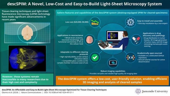
Three-dimensional (3D) imaging of organs and tissues is vital as it can provide important structural information at the cellular level. 3D imaging enables the accurate visualization of tissues and also helps in the identification of pathological conditions. However, achieving successful 3D imaging necessitates specific prerequisites, including the preparation of ‘cleared’ tissue samples—biological specimens rendered transparent by removing light-scattering components like lipids to enable comprehensive visualization of internal structures—utilization of fluorophores and adoption of an appropriate imaging technique. Selective plane illumination microscopy (SPIM) is a type of light sheet fluorescence microscopy (LSFM), which is one such 3D imaging technique employing a thin sheet of light to illuminate cleared tissues.
Light Sheet Fluorescence Microscopy Beneficial but Expensive
Significant advancements in tissue clearing techniques have aided their extensive use in biomedical imaging. However, LSFM systems have faced accessibility challenges due to high costs and complexity. To address this issue, a team of researchers from Juntendo University in Japan, comprising Associate Professor Kohei Otomo, PhD; Takaki Omura; and Professor Etsuo A. Susaki, MD, PhD, developed an innovative, low-cost LSFM called descSPIM – desktop-equipped SPIM for cleared specimens. They optimized the design of the microscope for rapid imaging of cleared tissue samples and verified its utility as an imaging tool using animal studies. Their findings were published Nature Communications.
Japanese Researchers Solve the Cost Barrier
Explaining the motivation behind the development of descSPIM, corresponding author, Susaki says, “LSFM remain prohibitively expensive for many end users, despite advancements in tissue clearing technologies. When I recognized this bottleneck, I wanted to develop an affordable and sufficiently performing light-sheet microscopes to enhance accessibility in biomedical research.” The team developed the descSPIM microscopic system with a focus on cost reduction, bringing the price down to approximately between $20,000 and $50,000. They also employed a simplistic design that requires less expertise for both assembly and operation.
The researchers also incorporated numerous unique features like utilizing a glass cuvette to replace the medium chamber and a two-stage synchronization process during imaging. Additionally, the mechanism of capturing optical data and converting them into images was streamlined, which resulted in a significant decrease of the overall time required for imaging.
Application Validated
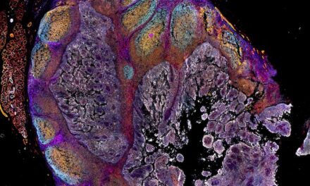
Further, the researchers validated the applications of descSPIM for 3D visualization of multiple cleared tissues and organs. descSPIM successfully imaged volumetric whole-brain samples of transgenic mice, allowing visualization of neuronal structures and distributions with cellular resolution. In cancer cell line-derived xenograft (CDX) models, descSPIM enabled 3D imaging of entire tumor masses, facilitating visualization of drug distribution within the tumor tissues. Furthermore, descSPIM was used to generate 3D images of thick tumor sections stained with fluorescent dyes, mimicking standard hematoxylin-eosin (HE) staining. While the resolution for clinical pathology examination was relatively low, it demonstrated the potential of descSPIM for future diagnostic applications.
The team open-sourced the design of descSPIM to facilitate extensive academic dissemination. “Commercial microscopy systems are generally designed as black boxes, where the internal mechanism is unknown to the users. However, the accessible design and open-source nature of descSPIM represent a departure from this norm. Its user-friendly construction and open-sourced nature foster a collaborative environment conducive to the development of pioneering imaging technologies,” explains Otomo, the first author of the study.
Light Sheet Fluorescence Microscopy Embraced Globally
Researchers globally have embraced the descSPIM system for a myriad of research endeavors, spanning from the detailed imaging and visualization of various organs, including the brain, hypothalamus, stomach, and intestine in mice and rats, to investigating cancer metastasis in the lungs. These diverse applications underscore the versatility and effectiveness of descSPIM across different biological contexts. Delving into the implications of their findings Otomo says, “When compared to commercially available light-sheet microscopes, our proposed DIY system is more than 10 times less expensive and strongly supports the dissemination of the tissue clearing and 3D imaging technology”.
The low-cost, simplistic yet innovative design of descSPIM light-sheet microscopy system has the potential to accelerate scientific research and result in path-breaking biomedical discoveries.
Featured Image: descSPIM enables three-dimensional imaging of diverse tissues like neural cells and cancerous tumors. The widespread dissemination and adoption of descSPIM can accelerate biomedical discoveries. Image: Etsuo A. Susaki from Juntendo University

