Olympus’ third annual Global Image of the Year Life Science Light Microscopy Award is now open for entries through Jan. 31, 2022. Each year, the competition recognizes the best in life science imaging worldwide to inspire and showcase art through microscopy.
Contestants may enter by uploading up to three images, with a description of the equipment used, at the Olympus website. Winners will be selected by a jury and announced in April 2022.
Global Image of the Year Contest Details
Prizes include an Olympus SZX7 stereo microscope with a DP28 digital camera for the global winner and an Olympus CX23 upright microscope for the regional winners in Asia, Europe, and the Americas.
The international jury includes experts from both science and the arts, including Wendy Salmon, director of the Hooker Imaging Core Facility at the University of North Carolina at Chapel Hill School of Medicine; Geoff Williams, manager of the Leduc BioImaging Facility at Brown University; Harini Sreenivasappa, microscopy facility manager of the Cell Imaging Center at Drexel University; Rachid Rezgui, research instrumentation scientist at New York University Abu Dhabi; Siân Culley, postdoctoral research associate at the MRC Laboratory for Molecular Cell Biology at UCL; Stefan Terjung, operational manager of the Advanced Light Microscopy Facility at EMBL Heidelberg; Wen-Biao Gan, director of the Institute of Neurological and Psychiatric Disorders at Shenzhen Bay Laboratory; and Anne Beghin, research assistant professor at MechanoBiology Institute, National University of Singapore.
All entries will be evaluated based on artistic and visual aspects, scientific impact, and microscope proficiency.
More information about the Global Image of the Year Life Science Light Microscopy Award, including jury members’ biographies, last year’s winning images, and the full terms and conditions, can be found at the Olympus website.
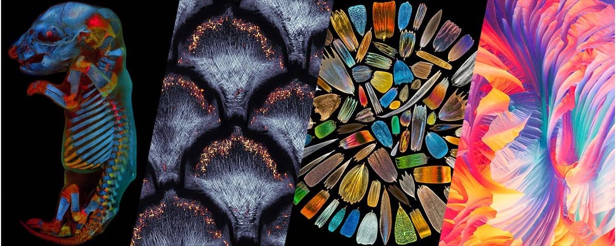
Featured Image: A collage featuring the 2020 Global Image of the Year Life Science Light Microscopy Award winners. From left: Image of the Year 2020 – The Global Winner, Werner Zuschratter (Germany), Whole rat embryo three channel large scale confocal image of a fixed and cleared rat embryo. Two channels show different autofluorescence sources of the tissue, whereas the third channel shows the skeleton stained by alizarin red; Image of the Year 2020 – Regional Winner (Europe, the Middle East and Africa), Grigorii Timin (Switzerland), Collagen fibers (second harmonic generation) and dermal pigment cells (autofluorescence) in African house snake embryonic skin; maximal intensity projection of 10 confocal slices; Image of the Year 2020 – Regional Winner (Asia-Pacific), XinPei Zhang (China), Scales collected from the wings of over 40 species of butterflies were photographed individually and finally assembled into this image; Image of the Year 2020 – Regional Winner (Americas), Justin Zoll (U.S.A.),Polarized light microscopy panorama of l glutamine and beta alanine crystals. Photo: Olympus


