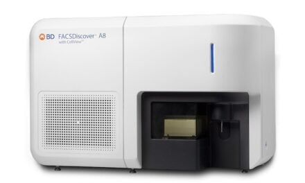
NEW YORK (Reuters Health) – Use of the ThinPrep Imager, a computerized system that is applied to liquid based cytology, can improve the detection of cervical squamous disease and intraepithelial neoplasia, according to a report in the June 28th issue of BMJ Online First.
The findings stem from a comparison of the imager with manually read conventional cytology. A total of 55,164 split sample pairs from consecutive patients were analyzed.
The rate of unsatisfactory slides with imager-read cytology was 1.8% compared with 3.1% with conventional cytology (p < 0.001), lead author Dr. Elizabeth Davey, from the University of Sydney in Australia, and colleagues note.
Imager read cytology was also more likely to classify slides as abnormal: 7.4% vs. 6.0% overall and 2.8% vs. 2.2% for cervical intraepithelial neoplasia (CIN) grade 1 or higher.
Compared with conventional cytology, imager-read cytology detected 71 additional cases of high grade histology. Using CIN grade 1 as the threshold for colposcopy referral, the imager detected 1.29 more cases of high grade squamous disease per 1000 women screened than did conventional cytology, the report indicates.
The authors note that the improved detection of CIN grade 2 lesions seen with the ThinPrep Imager might allow for longer intervals between screenings. Conversely, the increased detection of low grade lesions might increase rates of further testing.
BMJ Online First 2007.


