Summary
Cambridge researchers demonstrated that AI could accurately identify drug-resistant infections from microscopy images, significantly reducing diagnostic time.
Takeaways
- AI Identification: An AI algorithm can be trained to identify drug-resistant bacteria from microscopy images without testing against drugs.
- Rapid Diagnosis: The AI approach reduces the time needed to identify drug resistance from several days to just a few hours.
- Potential and Limitations: While the method shows promise in combating antimicrobial resistance, practical deployment and cost-effectiveness remain challenges.
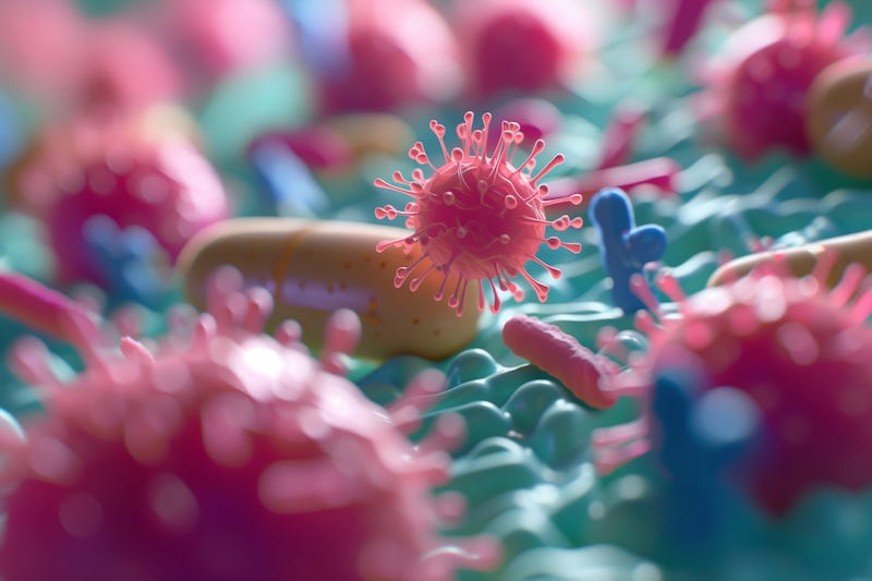
Artificial intelligence (AI) could be used to identify drug resistant infections, significantly reducing the time taken for a correct diagnosis, Cambridge researchers have shown. The team showed that an algorithm could be trained to identify drug-resistant bacteria correctly from microscopy images alone.
Antimicrobial Resistance: a Global Health Issue
Antimicrobial resistance is an increasing global health issue that means many infections are becoming difficult to treat, with fewer treatment options available. It even raises the spectre of some infections becoming untreatable in the near future.
One of the challenges facing healthcare workers is the ability to distinguish rapidly between organisms that can be treated with first-line drugs and those that are resistant to treatment. Conventional testing can take several days, requiring bacteria to be cultured, tested against various antimicrobial treatments, and analysed by a laboratory technician or by machine. This delay often results in patients being treated with an inappropriate drug, which can lead to more serious outcomes and, potentially, further drive drug resistance.
In research published in Nature Communications, a team led by researchers in Professor Stephen Baker’s Lab at the University of Cambridge developed a machine-learning tool capable of identifying from microscopy images Salmonella Typhimurium bacteria that are resistant to the first-line antibiotic ciprofloxacin—even without testing the bacteria against the drug.
S. Typhimurium causes gastrointestinal illness and typhoid-like illness in severe cases, whose symptoms include fever, fatigue, headache, nausea, abdominal pain, and constipation or diarrhea. In severe cases, it can be life threatening. While infections can be treated with antibiotics, the bacteria are becoming increasingly resistant to a number of antibiotics, making treatment more complicated.
The team used high-resolution microscopy to examine S. Typhimurium isolates exposed to increasing concentrations of ciprofloxacin and identified the five most important imaging features for distinguishing between resistant and susceptible isolates.
They then trained and tested machine-learning algorithm to recognise these features using imaging data from 16 samples.
Algorithm Correctly IDs Disease in Microscopy Images
The algorithm was able to correctly predict in each case whether or not bacteria were susceptible or resistant to ciprofloxacin without the need for the bacteria to be exposed to the drug. This was the case for isolates cultured for just six hours, compared to the usual 24 hours to culture a sample in the presence of antibiotic.
“S. Typhimurium bacteria that are resistant to ciprofloxacin have several notable differences to those still susceptible to the antibiotic. While an expert human operator might be able to identify some of these, on their own they wouldn’t be enough to confidently distinguish resistant and susceptible bacteria,” says Tuan-Anh Tran, PhD, who worked on this research while a PhD student at the University of Oxford and is now based at the University of Cambridge. “The beauty of the machine learning model is that it can identify resistant bacteria based on a few subtle features on microscopy images that human eyes cannot detect.”
In order for a sample to be analyzed using this approach, it would still be necessary to isolate the bacteria from a sample—for example a blood, urine or stool sample. However, because the bacteria do not need to be tested against ciprofloxacin, this means the whole process could be reduced from several days to a matter of hours.
AI and Microscopy Images: An Imperfect Match—for Now
While there are limitations to how practical and cost effective this particular approach would be, the team says it demonstrates in principle how powerful artificial intelligence could be in helping the fight against antimicrobial resistance.
“Given that this approach uses single cell resolution imaging, it isn’t yet a solution that could be readily deployed everywhere. But it shows real promise that by capturing just a few parameters about the shape and structure of the bacteria, it can give us enough information to predict drug resistance with relative ease,” Sushmita Sridhar, PhD, who initiated this project while a PhD student in the Department of Medicine at the University of Cambridge and is now a postdoc at the University of New Mexico and Harvard School of Public Health.
The team now aims to work on larger collections of bacteria to create a more robust experimental set that could speed up the identification process even more and allow them to identify resistance to ciprofloxacin and other antibiotics in a number of different species of bacteria.
“What would be really important, particularly for a clinical context, would be to be able to take a complex sample—for example blood or urine or sputum—and identify susceptibility and resistance directly from that,” says Sridhar. “That’s a much more complicated problem and one that really hasn’t been solved at all, even in clinical diagnostics in a hospital. If we could find a way of doing this, we could reduce the time taken to identify drug resistance and at a much lower cost. That could be truly transformative.”
The research was funded by Wellcome.

