Researchers developed a stain-free system using silicon slides to reduce tissue preparation time and support artificial intelligence analysis.
Scientists at King Abdullah University of Science and Technology (KAUST) developed a new stain-free imaging platform designed to analyze tissue samples more quickly and consistently. The research, published in Advanced Science, aims to support future artificial intelligence (AI)-assisted cancer diagnostics.
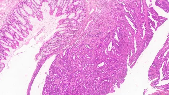
The platform uses engineered silicon slides to generate detailed structural color images directly from tissue samples, removing the need for conventional chemical dyes. While chemical staining is widely used in pathology laboratories, the process can add time to diagnostic workflows and vary depending on laboratory conditions and preparation methods.
In a study evaluating tissue samples from 120 patients, the platform achieved a 99% agreement rate with conventional pathology assessments for colorectal tissue. Pathologists reached the same diagnostic conclusions in almost all cases while using the faster, stain-free process.
Improving Workflow Efficiency
Because the method eliminates chemical staining, researchers observed a reduction in preparation time compared with conventional workflows. Early results indicate the process could reduce sample preparation time by approximately 40% to 50%, while improving consistency by removing variability linked to staining conditions.
“This research focuses on improving one of the most important steps in diagnosis: how tissue samples are prepared and reviewed,” says Qiaoqiang Gan, professor of material science and engineering at KAUST, in a release. “Traditional staining methods can be influenced by preparation steps, reagent quality, and laboratory conditions. By generating consistent digital images without dyes, we can reduce variability and create data that is more reliable for both clinical review and future AI-assisted analysis.”
Broad Clinical Applications
The platform was first validated using colorectal tissue samples, a major health priority in Saudi Arabia. However, researchers also tested the technology on breast, lung, and thyroid tissue samples. The platform captured key histological features comparable to those found on slides prepared with conventional stains.
The research team is now working with clinical partners, including King Faisal Specialist Hospital & Research Centre Madinah, to evaluate the platform across broader healthcare settings. The researchers are also working to assess pathways for future clinical and commercial use of the system.
Photo caption: Healthy and cancerous regions in results
Photo credit: KAUST

