

In 1990, the level of penetration of digital radiology in the United States was about 2%; in 2000, it was 90%, according to Ajit Singh, PhD, president and CEO of BioImagene in Sunnyvale, Calif. “So it took about 10 years to go from nearly zero to nearly 100. I believe [the adoption] of digital pathology will be a little bit faster—more than 5 years, less than 10—because we now have all the experiential learning from radiology,” Singh says.
Currently, however, digital pathology is just beginning to penetrate the clinical laboratory market, although it is well entrenched in medical education and nearly mainstream in research. “Every medical school, every vet school in the world is using digital technology. It’s so much better to teach a roomful of students with digital than it is to give them all microscope slides. It saves money, and everyone can look at the same thing,” says Ole Eichhorn, chief technology officer of Aperio, located in Vista, Calif.
Clinical pathology has been slow to adopt, in part because of the early generation of the technology as well as the natural human resistance to change. “There has been a lot of controversial discussion about where this fits. Pathologists can see it used in terms of helping them in tricky situations or quantifying data when they’ve got large volumes, but there are also those who are concerned about the impact on pathology: If it does the diagnosis for you, it may be considered a crutch or thought to replace the pathologist,” says Rhonda Henshall-Powell, PhD, product manager with Biocare Medical in Concord, Calif.
As a result, the use of digital systems is still in its initial stages, slowly penetrating through niche applications. “There are 200 million glass slides read in the United States every year. My estimate would be that no more than two million are read digitally today. That’s 1 percent, but for immunohistochemistry, it could already be about 20 percent,” Singh says.
Those in pathology foresee digital systems as the future of the field, with estimates of mainstream adoption ranging from 2 to 10 years. “In some applications and diagnostic tasks they will become mainstream, while in others manual eye-assessment will remain the governing routine. The distinction between the two will be dictated by the benefit the patient and pathologists get by the use of tools—such as CAD—in terms of money savings, better treatment, and earlier detection,” says Tsafrir Kolatt, PhD, business development and program director for Applied Spectral Imaging, Vista, Calif.
Racing to Improve
“The more digital systems can increase their capabilities and the ability to read, over-read, or do consultations, it will promote usage and therefore push for adoption,” says Ravi Sharma, CEO of 4medica in Culver City, Calif.
Singh notes three dimensions along which these hardware and software technologies have evolved: functionality, usability, and speed. With each new generation, these improve, and many feel they have finally reached an acceptable level.
Digital pathology hardware consists of the devices used for scanning. “The main features of these instruments are how fast they are and the resolution of the images generated. So there is a bit of an arms race between vendors to keep making faster scanners,” Eichhorn says. He expects that, someday, slides will be scanned in 10 seconds, but currently, it takes about 1 to 2 minutes. Most companies offer autoloaders: scanners that can be loaded with multiple slides and operate independently of a technologist.
Higher-resolution scanners take longer to capture an image and are currently often reserved for niche applications. Specialized equipment is also available for fluorescence.
On the software side, basic functionality focuses on the digitization and manipulation of scanned images. Advanced applications include the identification of abnormalities, counting of events and objects, classifying event and objects, IHC-scoring, cytology sample evaluation, tuberculosis evaluation, FISH classification, and tissue characterization, Kolatt lists.
“People are building new applications every day, and some of them are actually driving adoption,” Eichhorn says. Other companies are focused on adding workflow management to a system’s functionality.
“Once the stack of trays is effectively removed from the pathologist’s desk because those slides are digitized, pathologists still need a system that brings their entire caseload together into a consolidated unit they can organize, triage, prioritize, and filter, which has to happen for enterprise adoption of digital pathology,” says Tony Melanson, vice president of strategy and marketing at Omnyx in Pittsburgh.
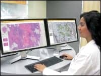
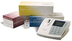
BioImagene’s Virtuoso is a Web-based digital pathology solution that allows users to view, manage, archive, and analyze digital images.
Automating and Redefining Care Standards
Yet even though these capabilities are undergoing refinement, there is already value in the use of digital systems. “I can summarize the benefits in two categories: one, it gives you the ability to do what you already did, albeit more efficiently, which I call automating the standard of care; and two, it offers the ability to do things you simply could not do before—I call that redefining a standard of care,” Singh says.
Automating the standard of care means improvements in the speed and quality of existing applications that translate into a more accurate and rapid diagnostic. Slides no longer need to be physically sent to the appropriate pathologist for diagnoses, consultations, or second opinions but can be shared electronically, reducing time and cost and enabling increased volume.
“To get the right specialist to look at a slide is a logistical challenge, and people are always mailing slides around. So by having a digital image, it makes it possible for anyone to view it anywhere at any time,” Eichhorn says.
Storage and retrieval become easier as well. Rather than a “Raiders of the Lost Ark warehouse effect of slide storage,” as Eichhorn colorfully compares it, slides are always locatable and retrievable. They do not get lost, fade, break, or disappear. Digital images also make it easier to redefine the standard of care. The software applications are designed to increase the pathologist’s ability to make a confident diagnosis by removing the subjective nature of the results. “There are immunohistochemical assays that people use to measure the type of cancer you have. The pathologist looks at the slides and determines what percentage of the cells are stained, making an estimate, say ‘about 30 percent.’ But a computer can measure that to the fifteenth decimal place, get it absolutely right, and be the same answer that any pathologist has anywhere,” Eichhorn says.
Comparison between slides also becomes easier and, ideally, patient outcomes improve. “We’ve demonstrated that with digital pathology, you can bring that false positive for [HER2 in breast cancer patients] from 18 percent to under 1 percent,” Singh says.
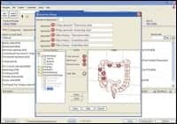
Path4 enables physicians to use a drag-and-drop functionality to identify biopsy locations on organ images. The system automatically generates labels with description of the locations.
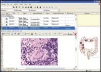
PDF reports, with embedded light microscope images, can be viewed in 4medica’s Path4 solution, along with the original order’s organ image with the identified biopsy locations.
Obstacles to Adoption
However, despite the many benefits, there have been some limitations that have affected adoption, including a lack of broad-based applicability; limited functionality, including workflow management capabilities; integration with other health information databases, such as the EMR and LIS; and storage, which is massive.
“There are a wide variety of sample types in pathology—different kinds of tissue, different preparations, different stains—and some of them are more amenable to being scanned and looked at digitally than others,” Eichhorn says. When pathologists find the digital image inadequate (and laboratories should note the rescan rate of their scanning systems for an indication of performance, Eichhorn notes), they turn to the glass slide.
Speed, quality, and functionality have vastly improved since the first generation of digital pathology platforms, but they do leave room for improvement before wide-scale adoption. Pathologists need to be comfortable with all aspects of the system so that they are comfortable with the workflow and diagnosis.
Melanson notes speed is reaching the point of acceptability, while image quality is likely to undergo greater scrutiny. “People are going to very critically evaluate image quality because these systems have not yet been FDA approved for broad diagnostic use,” Melanson says.
But limitations to workflow may be a bigger obstacle to greater adoption. “With a case-driven workflow, pathologists have to go into folders and open individual images to view them. So until you can get all of their workflow into a single system with high enough quality, these digital tools, though interesting, tend to be distractions,” Melanson says.
This means the systems will need to integrate with other information databases. “To view an image in isolation from the rest of the clinical data is not very useful,” Singh says. So pathologists need access to data in other systems, such as the EMR, the LIS, and the RIS, to both retrieve and save information.
Fortunately, manufacturers have realized this already. “Several of these interfaces already exist or can be rapidly developed,” says Wally Soufi, CEO of Novovision Inc, located in Princeton, NJ.
Ease of use is particularly key since pathologists tend to be a conservative group. Digital pathology has been on the scene for about 10 years; light microscopes have been used for hundreds. “Pathologists are making life-and-death decisions, and that responsibility weighs on them. When you have a 60-year-old pathologist who has been looking at cases for 40 years, getting him or her to change the way they do things is hard,” Eichhorn says.
Usability
Fortunately, training users of these systems is relatively easy. For laboratory technologists, who are accustomed to working regularly with instrumentation, training is fairly straightforward and varies according to the vendor, running anywhere from a few hours to a few days. Generally, manufacturers seek to make the devices as easy to use as possible to keep errors low and productivity high.
Pathologists require about as much time as the histotechnicians but often need to start with the basics since the use of computers in their labs is fairly new. “In a lot of cases, this might be the first time the physician has really used a computer,” Eichhorn says.
Subsequently, manufacturers seek to create as intuitive an interface as possible and capture pathologists’ attention at the right time. Some have implemented a training process that imparts the iScan Coreo Au is BioImagene’s second-generation brightfield scanner with 160-slide capacity. basics first and greater functionality a month or so later. “It isn’t until they actually try to use it that they are really open to being trained,” Eichhorn says.
They also require reliability before committing to a system. “Once you put a scanner into a high-throughput lab, it needs to be designed for constant operation,” Melanson says. Frequent breakdowns will impede confidence and use.
Currently, some manufacturers have found the meantime to failure to be about 1 year. The problem often originates within the slide-moving components, which have to handle slides of varying size and shape. However, most manufacturers are looking to improve reliability.
Increased reliability will add to the value of the system, which currently offers a return on investment through tangibles, such as saved shipping and increased consultation revenue, as well as intangibles, such as greater efficiencies and more accurate diagnoses.
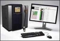
iScan Coreo Au is BioImagene’s second-generation brightfield scanner with 160-slide capacity.
Today’s digital pathology devices and systems range from $15,000 to $300,000 and may be available as a capital purchase or with a pay-per-use approach. High-quality monitors for remote pathologists and storage can be additional expenses but are far less prohibitive than when radiology debuted digital images. However, because pathology images are much larger than those captured in radiology—by about two orders of magnitude, according to Eichhorn—more storage is needed.
In addition, regulations continue to require the glass slides be saved, but since they are no longer needed for clinical use, they can be stored off-site, saving hospital space. Reimbursement will also need to catch up. “There are CPT codes for specific procedures done with digital pathology, but there aren’t many of them. The ones that do exist will certainly be one of the reasons people will adopt,” Eichhorn says.
Another will be increased collaboration among pathology providers. For instance, Biocare does not currently offer digital capabilities but is looking to collaborate with a digital provider to partner its stains.
“With the integration of computer tools for image analysis and enhancements, computer-aided diagnosis, and the integration of anatomic pathology information systems, it is likely the scanning of whole slides will contribute to health care cost-reduction initiatives, as well as improve patient care,” Soufi says. Just ask a radiologist.
Renee Diiulio is a contributing writer for CLP.

