Respiratory viruses are commonplace, from the common cold to influenza to pneumonia. The Centers for Disease Control and Prevention (CDC) suggests that most children will have serologic evidence of respiratory syncytial virus (RSV) by the age of 2 years.
Although not often fatal, these conditions are more dangerous, as well as more prevalent, among the very young and the very old. And their misery is opportunistic—easily contagious, many can spread through a mere sneeze.
Antiviral medications can help, but only if administered right away. Two of the influenza antiviral medications approved by the FDA—oseltamivir (Tamiflu) and zanamivir (Relenza)—can decrease illness severity and length, but they must both be started within 48 hours of the onset of symptoms to have a positive effect on outcome. Antibiotics, naturally, do not help to alleviate viral symptoms or eliminate the disease, and can contribute to drug-resistance problems if improperly prescribed.
It is, therefore, important for physicians to make informed choices when treating patients with respiratory viruses, particularly those who may demand medications. Test results provide some of the best information, detecting and differentiating conditions and helping to clarify a treatment protocol.
Rapid test results can improve the outcome of not only an individual, but also the community at large since early treatment can halt the spread of the disease. Laboratories have therefore traditionally been challenged to produce results as quickly as possible, a demand that has not necessarily lessened with the advent of point-of-care tests.
Conventional Cultures
While physicians can use a rapid test to help determine a course of treatment, the results are qualitative. More sophisticated tests are often used to confirm results as well as track infections and identify outbreaks. Cultures have traditionally been the gold standard, but their lengthy turnaround is a distinct disadvantage.
ARUP Laboratories, Salt Lake City, notes that most of its agents require 24 to 72 hours to culture a specimen.1 Other conventional cultures can take as long as 30 days.
Shell vial cultures tend to be on the shorter end of the scale, and in some laboratories they have replaced traditional culture methods. Shih et al conclude that the technique rapidly detects and identifies respiratory tract viruses.2 In comparing shell vial cultures to conventional techniques, they found the shell vial culture/immunofluorescent staining method allowed detection of 81.5% of influenza A virus, 72% of parainfluenza virus, 82.6% of RSV, and 79.6% of adenovirus in 24 hours versus the 3 to 30 days needed for traditional cultures.2
The shell vial methods use a centrifuge to optimize the assay, sometimes producing more accurate results along with faster ones. Meqdam and Nasrallah found that a shell vial culture assay showed the highest sensitivity and specificity in the detection of RSV when compared with a conventional culture assay and a direct immunofluorescence assay.3
Direct Fluorescence
Direct immunofluorescence assays, however, can provide an excellent option for diagnostics, particularly considering their faster turnaround times and reduced manual labor. Cultures may have traditionally been the gold standard, but some could say that standard has begun to tarnish. Faster diagnosis leads to faster treatment and, in some instances, faster recovery. Direct fluorescence assays are faster than both cultures and real-time PCR (polymerase chain reaction).
A team comprised of researchers from the Memorial Medical Center in Springfield, Ill, and the internal medicine department of the Southern Illinois University School of Medicine, also in Springfield, compared patients diagnosed through standard techniques only (such as enzyme immunoassays, shell vial assays, and culture tube assays) with those whose samples were also screened with immunofluorescent testing (FA) directly on cytocentrifuged samples.4 The rapid turnaround resulted in more positive outcomes.
Patients who had been screened with the additional test had faster result turnarounds (0.9 days versus 4.5 days), shorter hospital stays (5.3 days versus 10.6 days), and fewer related expenses ($2,177 versus $7,893).1 The researchers concluded that, “The cytospin FA markedly decreased turnaround time and was associated with decreased mortality, length of stay, and costs, and with better antibiotic stewardship.”4
Tests such as the PathoDX Respiratory Virus Panel from Remel Products/Thermo Fisher Scientific,Lenexa,Kan,and Bartels RSV Monoclonal DFA (Direct Fluorescent Antibody) Kit from Trinity Biotech PLC, Berkeley Heights, NJ, use direct fluorescence to detect respiratory diseases. Bartels RSV Monoclonal DFA targets RSV only, while the PathoDX RVP screens for seven diseases: RSV; influenza A and influenza B; parainfluenza viruses 1, 2, and 3; and adenovirus. Both use monoclonal antibodies. Bartels RSV labels its monoclonal antibodies with fluorescein; PathoDX uses FITC-labeled mouse monoclonal antibodies specific to each virus.
These tests can provide clinically valuable data more quickly than a culture,but they may be less sensitive. In addition, adequacy of the specimen can impact accuracy.
 |
| A variety of tests have become available to help laboratorians determine the sources of respiratory infections. |
According to ARUP, “the sensitivity of direct fluorescent antibody testing for influenza, parainfluenza, and respiratory adenovirus improves with specimen type (eg, true sputum and nasopharyngeal are better than throat swabs).”1 The respirator secretions in the sample must contain intact respiratory epithelium to make it adequate.1 False negatives can result with too few cells on the slide or cell clumping1; false positives may occur in samples with excess mucus or a lumpy smear by “nonspecifically trapping the antibody reagent.”1
Technique and technology can help to ensure adequate specimens. A study conducted by Landry and Ferguson found only 7.5% of samples were inadequate for direct fluorescence antibody staining after preparing the slides with cytospin.5 Even so, direct fluorescence has failed to replace culture as the gold standard.
Real-Time PCR
Real-time PCR comes closer. Though slower and more expensive than direct fluorescent methods, PCR has frequently been shown to be more sensitive. A study published earlier this year compared real-time PCR, immunofluorescence, and viral isolation in the detection of RSV in the nasopharyngeal aspirate of infants (up to 2 years),and found real-time PCR was the most sensitive.6 An earlier study that looked at real-time PCR versus conventional methods in the diagnosis of lower respiratory tract infections in children also found real-time PCR to be the more sensitive methodology, increasing the diagnostic yield twofold.7
Additional benefits include the fact that real-time PCR technology can be easily integrated into the laboratory workflow while requiring little hands-on time. The technology is fairly easy to use; it reduces the risk of cross-contamination; and it permits the detection of multiple viruses at once, including multiplex (two to seven targets) and massively multiplexed (10 to 20 targets).
The Multiplex RT-PCR ProFlu+ Assay from Prodesse Inc, Waukesha, Wis, was one of the first multiplex real-time PCR tests to be approved by the FDA. It also is the first infectious disease assay to be approved to run on the NucliSENS easyMAG, the automated nucleic acid extraction system from Durham, NC-based bioMérieux Inc.
The diagnostic simultaneously detects and differentiates influenza A and B and RSV in about 3 hours, still much quicker than conventional methods. In addition, the FDA has removed its recommendation that negative results for influenza A or influenza B be confirmed by culture—a first for a molecular test, according to the company.
In a company press release, Prodesse CEO Tom Shannon states, “Because culture is considered the ‘gold standard’ for respiratory viruses, only a test that shows exceptional performance compared to culture can have this recommendation removed. Our clinical trial sites detected 46 percent more positives than culture when using our ProFlu+ Assay. Further testing by genetic sequencing determined that nearly 90 percent of the 103 discrepant samples were likely true positives.”
In a poster presented by Johns Hopkins at the 2008 Clinical Virology Symposium, researchers found that the diagnostic yield increased slightly with ProFlu+ when compared to immunochromatography in the diagnosis of RSV, influenza A, and influenza B in nasopharyngeal aspirates.8
Prodesse expects to expand this viral testing line with two respiratory virus diagnostics and a third enteric virus test in the pipeline. Pro hMPV+ is currently in clinical trials for the detection of human metapneumovirus. ProParaflu+, a multiplex assay for differentiating parainfluenza viruses, and ProGastro Cd for C. difficile will begin clinical trials soon.
PCR Pipeline
Seegene Inc, Rockville, Md, is also working toward FDA approval for its multiplex real-time PCR tests, which are currently CE marked. The Seeplex 18-Plex Respiratory Test detects 11 RNA respiratory viruses, two DNA, and five bacterial infections; the Seeplex RV12 ACE Detection simultaneously detects 12 respiratory viruses; and the Seeplex RV5 ACE Screening enables rapid screening for the most prevalent respiratory viruses.
Multiplex screening is more efficient, labor-wise, time-wise, and cost-wise. “[With the Seeplex RV12 ACE Detection], laboratories get 12 different results for the price of one or two tests,” says Jong-Yoon Chun, PhD, Seegene’s CEO and inventor of the Seeplex platform. He estimates that a hospital laboratory with a patient volume of 1,000 patients per month would save $1 to $2 million annually.
The technology is based on a dual priming oligo, or DPO, platform that works in conjunction with automatic detection systems, such as capillary electrophoresis or sequencing instruments. The DPO-based primers are comprised of two priming parts that serve to amplify only the target gene, generating consistently high PCR specificity. Amplicons are separated and analyzed automatically. “Our goal is to provide a very accurate diagnostic for the proper treatment of the patient,” Chun says.
 |
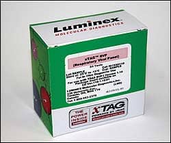
| The FDA-cleared xTAG Respiratory Viral Panel from Luminex detects and identifies 12 viruses and viral subtypes. |
Very accurate and very fast—because more than one disease marker can be detected at a time, results are produced rapidly. The average turnaround is about 5 hours, according to Chun.
Seegene is not the only company with multiplex real-time PCR tests in development. Nanogen Inc, San Diego, has been awarded a $10.4 million, 2-year contract from the CDC to develop a multi-analyte molecular diagnostic that will detect and differentiate influenza A, influenza B, seasonal flu (H1N1 and H3N2) strains, and RSV. The test will be developed in partnership with the Medical College of Wisconsin and HandyLab Inc.
The project is based on the company’s proprietary MGB probe technology, and is expected to produce an assay that can be completed in half the time it takes to run molecular diagnostics today while increasing sensitivity. Competition exists with not only similar technology but with tests that incorporate more complex technologies, such as microarrays and microspheres.
Multiplex PCR
The xTAG Respiratory Viral Panel, developed by Texas-based Luminex Corp, was the first multiplex test cleared by the FDA for infectious respiratory disease viruses. The diagnostic simultaneously detects and identifies 12 viruses and viral subtypes that together are responsible for more than 85% of RSVs.
The technology, based on the Luminex Universal Array, converts RNA into complementary DNA, which it then amplifies through PCR. DNA-specific labeled TAG primers are introduced to bind to viral targets; the resulting compounds bind to the complementary color-coded beads that are read by lasers of the Luminex xMAP instrument.
xTAG RVP is the first FDA-cleared assay for the detection and differentiation of influenza A subtypes H1 and H3 and the first FDA-cleared product to test for metapneumovirus, a virus discovered in 2001 that causes flu-like symptoms and is thought to be the second-leading cause of respiratory infection in children. Other viruses tested include influenza A; the subtypes H1 and H3; influenza B; parainfluenzas 1,2,and 3; and RSV. The test can provide qualitative results in a few hours.
“We recognized that current testing technology did not serve the patient very well because of the time involved,” says Jeremy Bridge-Cook, vice president of Luminex Molecular Diagnostics. “We also recognized the increase of a threat of pandemic flu as a public health issue.”
ViraCor Laboratories, Lee’s Summit, Mo, was the first national reference lab to offer the xTag from January to the end of June. Since the lab turns all patient results around in 24 hours, it did not offer viral testing.
“The xTAG is a major advancement in respiratory virus testing,” says Steve Kleiboeker, chief scientific officer and vice president of ViraCor. “Until now, clinicians have been playing a guessing game based largely on a patient’s symptoms. The high degree of sensitivity and specificity of xTAG helps eliminate incorrect diagnostics by greatly reducing the false negative results that are common in conventional testing methods.”
The population tested was largely post-transplant patients: 49.2% of the specimens tested were positive,and 5.5% of the specimens tested were dual infection, of which 75% contained rhinovirus. Triple infections observed were parainfluenza 3, rhinovirus, and RSV B; parainfluenza 3, rhinovirus, and adenovirus; and parainfluenza 3, rhinovirus, and metapneumovirus.
“We did not appreciate the burden of respiratory virus out there,” Kleiboeker says. “We are uncovering information that the physicians didn’t have before.”
Rapid Immunoassays
Rapid immunoassays also help to reveal clinically relevant data, on-site and often within 15 minutes. They enable a quick diagnosis and initiation of treatment with subsequent benefits of improved outcomes, reduced cost, and early control of the infection’s spread. For this reason, rapid antigen testing is one of the most popular methods for detecting respiratory viruses.
Rapid antigen detection includes enzyme immunoassays and fluorescent antibody stains. Newer tests seek to improve accuracy, which has not traditionally been extremely high in this category, though a reduction in subjective interpretation helps.
The QuickVue RSV by Quidel Corp, San Diego, indicates the presence of RSV with a pink/red test line. No line means no RSV. A blue control lines ensures the test is working properly. Results are revealed in 15 minutes with 92% sensitivity and 92% specificity when compared to cell culture.
The test requires only 30 seconds of hands-on time and one reagent. The CLIA-waived dipstick format works with a variety of sample types, including nasopharyngeal swabs, nasal or nasopharyngeal washes, and nasopharyngeal aspirates. “The QuickVue RSV test offers rapid, accurate detection with a choice of sample type, and its dipstick format is easy to use,” notes Tom Barman, senior marketing manager at Quidel.
The 3M Rapid Detection Flu A+B test, marketed by Response Biomedical Corp, Vancouver, BC, also produces fast results in the short time frame of 15 minutes. The test does not require subjective interpretation and features a rapid detection reader (the RAMP 200 Reader) that permits automatic recording and storage of test data as well as connectivity with the LIS.
“A busy lab can’t always spare the manpower required to stop and read a test with a short result window. The rapid detection reader gives the lab more flexibility in time and test management,” says Catherine Lathem, marketing manager for 3M Medical, St Paul, Minn.
The results give physicians more confidence in deciding on a treatment protocol, but it is recommended that negative results be confirmed by cell culture. The results may come too late to impact an individual’s outcome, but they can be useful by infection surveillance programs for the common good.
– Renee Diiulio is a contributing writer for CLP.
References
- ARUP Consult. Respiratory viruses: Indications for ordering. ARUP Laboratories. [removed]www.arupconsult.com/Topics/InfectiousDz/Viruses/RespiratoryViruses.html#[/removed]. Accessed August 11, 2008.
- Shih SR, Tsao KC, Ning HC, Huang YC, Lin TY. Diagnosis of respiratory tract viruses in 24 h by immunofluorescent staining of shell vial cultures containing Madin-Darby Canine Kidney (MDCK) cells. J Virol Methods. 1999;8(1-2):77-81.
- Meqdam MM, Nasrallah GK. Enhanced detection of respiratory syncytial virus by shell vial in children hospitalised with respiratory illnesses in Northern Jordan. J Med Virol. 2000;62(4):518-523.
- Barenfanger J, Drake C, Leon N, Mueller T, Troutt T. Clinical and financial benefits of rapid detection of respiratory viruses: an outcomes study. J Clin Microbiol. 2000;38(8):2824-2828.
- Landry ML, Ferguson D. SimulFluor respiratory screen for rapid detection of multiple respiratory viruses in clinical specimens by immunofluorescence staining. J Clin Microbiol. 2000;38(2):708-711.
- Reis AD, Fink MC, Machado CM, et al. Comparison of direct immunofluorescence, conventional cell culture and polymerase chain reaction techniques for detecting respiratory syncytial virus in nasopharyngeal aspirates from infants. Rev Inst Med Trop Sao Paulo. 2008;50(1):37-40.
- Van de Pol AC,Wolfe TFW, Jansen NJG, van Loon AM, Rossen JWA. Diagnostic value of real-time polymerase chain reaction to detect viruses in young children admitted to the paediatric intensive care unit with lower respiratory tract infection. Critical Care. 2006;10:R61.
- Gluck L,Wehrlin JA, Tilley C, Forman M, Valsamakis A. Utility of multiplex reverse transcription/real time PCR for detection of RSV, influenza A, and influenza B in nasopharyngeal aspirates. Poster by Johns Hopkins at the 2008 Clinical Virology Symposium. www.prodesse.com/USA/product/usIVD.html. Accessed August 11, 2008.


