A ground-breaking new eye scan could help identify autism in children years earlier than currently possible. The noninvasive technique utilizes a handheld device to find a pattern of subtle electrical signals in the retina that are different in children on the autism spectrum. The differences are directly linked to differences in brain development. The potential biomarkers for autism spectrum disorder could also allow for early detection of other disorders, such as attention deficit hyperactivity disorder.
The scan was tested on about 180 people with and without autism between the ages of 5 and 21 in collaboration with Yale University, University College London, and Great Ormond Street Hospital in London. Paul Constable, PhD, a senior lecturer in the College of Nursing and Health Sciences at Flinders University, has been searching for an autism biomarker since 2006, in an effort to improve early detection and intervention methods after his own child was diagnosed.
“The retina is an extension of the brain, made of neural tissue, and connected to the brain by the optic nerve, so it was an ideal place to look,” Constable says. “The test is quick and nonintrusive, and we anticipate it will be equally effective on younger children. Very early diagnosis means not only can children receive important interventions, but families are empowered to get the necessary supports in place, come to terms with the diagnosis, and make informed decisions.”
Reference
1. Constable PA, Ritvo ER, Ritvo AR. Light-adapted electroretinogram differences in autism spectrum disorder. J Autism Dev Disord. Epub ahead of print, February 7, 2020; doi: 10.1007/s10803-020-04396-5.
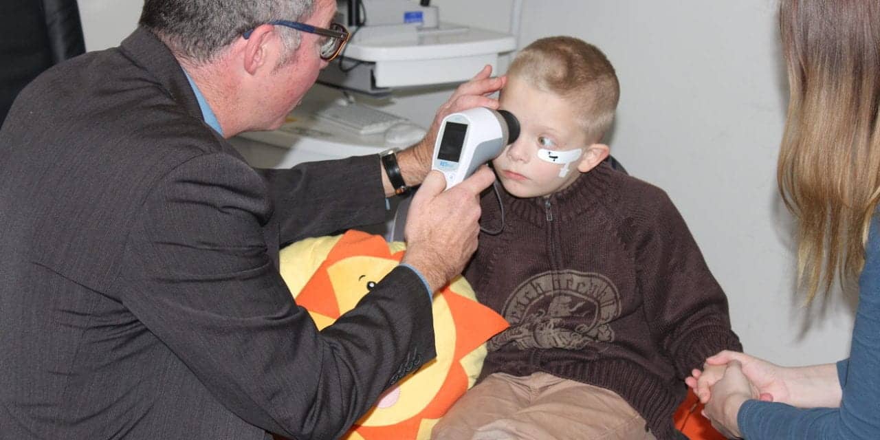
Featured image: Paul Constable, PhD, uses a handheld device to examine a child’s retina for signs of autism spectrum disorder. Photo courtesy Flinders University.


