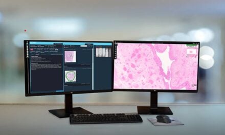
MD Anderson pathologist Savitri Krishnamurthy reflects on the evidence that helped bring digital pathology into clinical practice and the opportunities AI could create for patient care.
By Bryce Brown, director of new product enablement at Leica Biosystems
Nearly 20 years ago, Savitri Krishnamurthy, MD, saw a digital pathology scanner for the first time and began thinking about how the technology could change the way pathologists work.
Krishnamurthy, a professor of pathology at The University of Texas MD Anderson Cancer Center, has since helped build the evidence supporting digital pathology’s use in clinical practice. Speaking at a Leica Biosystems Open Day event, she discussed opportunities for digital imaging and artificial intelligence (AI) to improve how pathologists review tissue samples, collaborate with colleagues, and contribute to treatment decisions.
The shift comes as laboratories face growing demands. Global cancer cases are projected to increase from nearly 20 million in 2022 to 35 million by 2050, while the US could face a shortage of nearly 2,400 pathologists within the next decade.¹,² As laboratories look for ways to manage increasing workloads, digital pathology could play a larger role in how pathologists diagnose disease and support patient care.
Building the Evidence for a Digital-First Future
Although Krishnamurthy recognized the potential of digital pathology early on, she also understood that widespread adoption would take time.
“While many viewed the technology with skepticism, I saw its potential immediately. It was clear to me that digital pathology would reshape the field,” she said. “At the same time, my journey has reinforced an important lesson: Adopting truly disruptive technologies in healthcare is a marathon, not a sprint. It requires strong clinical evidence, sustained commitment, and champions willing to drive change.”
To help build that evidence, Krishnamurthy helped lead an early multi-institutional study comparing breast disease diagnoses made using conventional light microscopy and whole-slide digital imaging. Three breast pathologists from three medical centers independently reviewed 150 H&E-stained breast tissue slides, demonstrating that digital pathology achieved accuracy, precision, and reproducibility comparable to traditional microscopy.³ The findings supported its use for primary diagnosis and helped lay the groundwork for broader clinical adoption.
“Digital pathology has enabled me to evaluate tissue efficiently, conduct research more efficiently, and work faster than I ever could with glass slides alone,” she said.
Connecting Expertise Beyond the Microscope
For decades, pathology collaboration depended on moving glass slides between laboratories and institutions. Consultations, consensus reviews, and subspecialty input were constrained by transportation and logistics, introducing delays throughout the diagnostic process.
“People had to physically transport, file, and track them down to support collaboration and second opinions, which could get messy,” Krishnamurthy said. “With digital pathology, that barrier is removed. Pathologists can access images as soon as they are available from the lab, without waiting for slides to be physically delivered.”
Beyond removing geographic barriers, digital pathology can make it easier for pathologists to reach a consensus on difficult cases.
“Anatomic pathology can be subjective, and morphology may be interpreted slightly differently by different people,” she said. “Bringing greater uniformity to the field is key. Not everyone is sitting in the same place where you can easily show or pass around a slide. Digital pathology makes it far easier to obtain consensus opinions than was possible with glass slides alone. That is a tremendous advantage because consensus is one of the most important ways we can improve uniformity, reproducibility, and precision.”
How Digital Pathology Is Changing Patient Care
Although pathologists rarely interact directly with patients, their work plays a critical role in diagnosis and treatment decisions. As cancer care becomes more complex and personalized, pathologists are increasingly called upon to interpret biomarker results, molecular findings, and other information that can help guide patient care and advance precision oncology.
“With algorithms applied to an H&E slide, we can begin to extract far more information that was previously inaccessible, such as which patients may do well with chemotherapy or other treatment options,” Krishnamurthy said. “There are many questions we can ask from a single H&E slide. Why wouldn’t we use that additional information if it can help clinicians manage patients more effectively? Digital pathology is what makes that possible. It’s not simply about improving workflow efficiency; it’s about unlocking new layers of clinically relevant information that can directly support patient care.”
The rise of companion diagnostics is further expanding the pathologist’s role in guiding treatment decisions. Krishnamurthy said the growing use of molecular diagnostics, flow cytometry, immunohistochemistry, and other testing methods means pathologists must interpret more information than ever before.
“Healthcare is increasingly focused on tailoring treatment to the individual patient, and pathologists are at the center of that transformation,” she said.
In a 2023 study published in JCO Clinical Cancer Informatics, Krishnamurthy and colleagues developed and validated a deep-learning model that used digitized H&E-stained tissue images to predict response to neoadjuvant chemotherapy in patients with triple-negative breast cancer.⁴ The study showed how AI can detect clinically meaningful patterns in standard pathology slides, creating new opportunities to inform treatment decisions and personalize care.
Krishnamurthy also sees opportunities for AI to reduce the time pathologists spend reviewing slides.
“A typical mastectomy case can involve around 25 slides, and when I am signing out 20 mastectomy cases, I could be evaluating hundreds of slides under a light microscope,” she said. “Instead, imagine having a well-validated AI algorithm that can quickly identify which slides contain tumors and help distinguish them from negative cases.”
By directing attention to the most relevant slides first, she said, AI could help pathologists review cases more efficiently.
The implications extend beyond diagnosis. Drug discovery and development can take years of research, validation, and regulatory review. Bringing a successful therapy to market can take nearly 14 years and up to $1 billion.⁵
“Digital pathology and AI are helping accelerate research and transform the correlative sciences that underpin clinical trials,” she said. “By enabling faster, more scalable analysis of complex data, these technologies can help streamline the pathway from discovery to drug development and regulatory review. Ultimately, that means promising therapies emerging from successful clinical trials may reach patients in need more quickly.”
The Challenge of Reimbursement
As digital pathology moves from early adoption to wider implementation, reimbursement remains a significant barrier. Laboratories must invest in scanners, software, storage infrastructure, validation, and workflow redesign, often years before realizing the full clinical and operational benefits. While technology has advanced rapidly, reimbursement models have not.
The College of American Pathologists and the American Medical Association CPT Editorial Panel established digital pathology add-on codes that recognize the work and resources required to digitize glass slides for primary diagnosis, including workflows that support AI-enabled analysis.⁶ However, these codes do not fully offset the costs of adopting digital pathology.
Krishnamurthy said reimbursement will become increasingly important as laboratories look to incorporate AI tools into their diagnostic workflows. These tools could help pathologists identify information that guides treatment decisions, but the costs of implementing the technology remain a concern.
“If reimbursement evolves alongside these tools, laboratories will be better positioned to adopt digital pathology and AI without carrying the full financial burden,” she said.
The Path Forward
Adopting digital pathology requires planning and investment. The transition involves coordination across pathology, laboratory operations, IT, and executive leadership, with careful consideration of infrastructure, workflows, and data management.
As digital pathology and AI become more widely used, pathologists will need to combine their clinical expertise with new technologies to help guide diagnosis and treatment decisions. Laboratories that invest in digital pathology will also be better positioned to manage growing workloads and take advantage of new AI tools as they become available.
“If we do not go primary digital, how are we going to apply all the new advances coming into the field—particularly AI algorithms that are poised to improve inter-observer consistency, reproducibility, precision, and efficiency?” Krishnamurthy said. “Every pathologist wants to do their very best for patients. Digital pathology gives us a foundation to use these technologies responsibly and effectively.”
References
- Bray F, Laversanne M, Sung H, et al. Global cancer statistics 2022: GLOBOCAN estimates of incidence and mortality worldwide for 36 cancers in 185 countries. CA Cancer J Clin. 2024;74(3):229-63.
- American Society for Clinical Pathology. ASCP deepens commitment to pathologist workforce development. ASCP News. 2025 Sep 11. Available at https://www.ascp.org/news/news-details/2025/09/11/ascp-deepens-commitment-to-pathologist-workforce-development
- Krishnamurthy S, Mathews K, McClure S, et al. Multi-institutional comparison of whole slide digital imaging and optical microscopy for interpretation of hematoxylin-eosin-stained breast tissue sections. Arch Pathol Lab Med. 2013;137(12):1733-9.
- Krishnamurthy S, Jain P, Tripathy D, et al. Predicting response of triple-negative breast cancer to neoadjuvant chemotherapy using a deep convolutional neural network-based artificial intelligence tool. JCO Clin Cancer Inform. 2023;7:e2200181.
- National Center for Advancing Translational Sciences. New therapeutic uses (NTU). National Institutes of Health. Published 2024 Nov 5. Available at https://ncats.nih.gov/research/research-activities/ntu
- College of American Pathologists. Digital pathology codes. Available at https://www.cap.org/advocacy/payment-and-reimbursement-policy/digital-pathology-codes/
About the author: Bryce Brown is a leader in digital pathology and molecular diagnostics, currently serving as director of product enablement, digital pathology at Leica Biosystems, a Danaher company. Brown has also worked as a cytogenetic technologist at the Mayo Clinic and the University of Minnesota, where he contributed to high-impact clinical diagnostics in hematological disorders. He holds a bachelor of science in molecular biology from Winona State University.
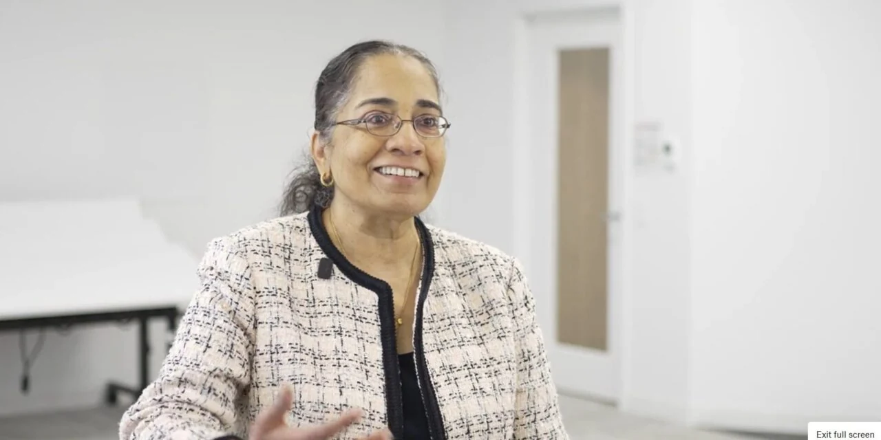
Photo caption: Savitri Krishnamurthy, MD
Photo credit: Leica Biosystems