
Analyzing the structure of existing biomarkers could reveal new information about cancer, helping laboratories deliver more accurate and clinically useful results.
By Arnon Chait, PhD, chief innovation officer, Cleveland Diagnostics
Cancer diagnostics has long focused on identifying biomarkers associated with disease and measuring how much is present in a patient sample. That approach could historically be traced to the advent of convenient technologies for measuring biomarker concentrations in circulation, rather than to any biological or scientific reasoning. The method has yielded some important observations and diagnostic tests, but for many cancers, biomarker concentration alone provides an incomplete picture.
An elevated biomarker concentration can reflect multiple biological processes, including inflammation, benign tissue changes, injury, or malignancy. This is one reason an abnormal result may identify a patient who needs further evaluation without clarifying whether the underlying disease is clinically meaningful. The next advancement in cancer detection may therefore come not from finding new biomarkers, but from learning how to obtain more relevant information from the biomarkers that laboratories already measure.
Looking Beyond Biomarker Concentration
Clinical laboratories routinely quantify proteins using immunoassays that are familiar, scalable, reproducible, and compatible with high-throughput testing. Yet quantitative results have an inherent limitation: They report abundance, sometimes related but not necessarily to the biological origin or disease relevance. A circulating protein can rise in response to inflammation, infection, benign tissue growth, trauma, or another condition. The laboratory may accurately measure that increased concentration without resolving its cause, potentially leading to additional imaging or invasive procedures.
Cancer compounds the problem because it is biologically heterogeneous and dynamic. Two patients with the same cancer label might have different molecular profiles, disease aggressiveness, clinical trajectories, and treatment needs. A useful diagnostic result must therefore do more than flag something abnormal. It needs to provide information that is analytically reliable, clinically interpretable, and actionable at the point of care.
Protein Structure Adds Biological Context
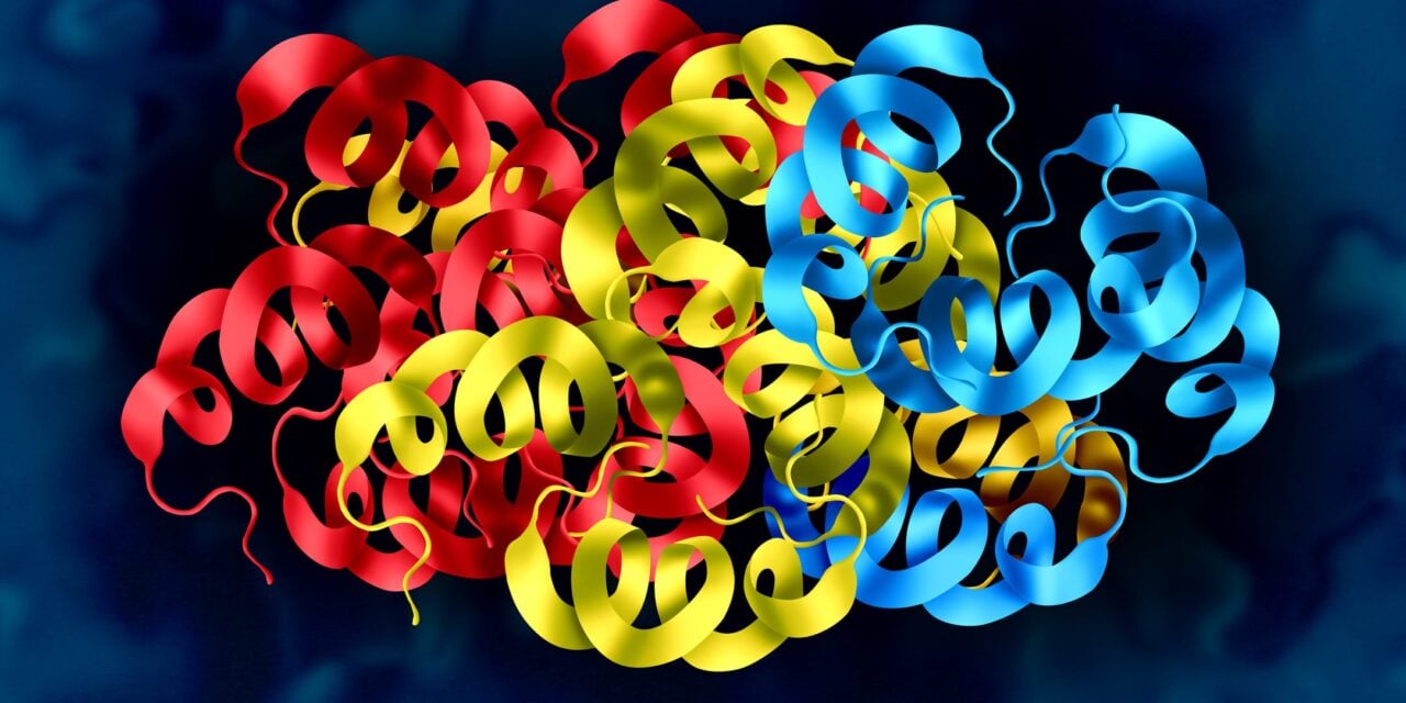
Proteins do not function merely because they are present. Their biological behavior depends on their structural characteristics, including how they fold, interact with other molecules, undergo post-translational modification, and exist in different molecular forms. Disease can alter those characteristics, meaning a protein released by a cancer cell can differ structurally from the same protein released by healthy or benign tissue, even if standard assays identify both as the same analyte. Detecting those structural variants could reveal whether a biomarker signal is more closely linked to an underlying malignant process.
This is conceptually different from simply measuring a biomarker with greater sensitivity. Highly sensitive assays and high-plex proteomic technologies offer important capabilities, from detecting low-abundance proteins to generating broad biological discovery data. But they address a different question from structural biomarker analysis, which explores whether a detected signal carries characteristics associated with the underlying disease process.
Prostate Cancer Illustrates the Opportunity
Prostate-specific antigen (PSA) is one of the best-known examples of both the value and the limitations of a concentration-based biomarker. PSA remains an important initial tool for screening and identifying patients who may require further evaluation. But PSA levels can increase for reasons other than cancer, including benign prostatic hyperplasia, inflammation, infection, and recent manipulation of the prostate. For that reason, an elevated PSA result does not directly answer the question that matters most to the patient and clinician: Is there a meaningful likelihood of clinically significant prostate cancer here?
Structural analysis of PSA provides one example of how an established biomarker can be evaluated differently. The US Food and Drug Administration-approved IsoPSA test assesses structural variants of PSA to aid in the detection of high-grade prostate cancer in patients with an elevated PSA who are being considered for biopsy. Rather than replacing PSA, it can add specificity to the diagnostic work-up after an initial elevated result.
This approach is not limited to prostate cancer or to a particular assay. A familiar biomarker can contain clinically relevant information beyond its measured concentration. If laboratories can access that information in a practical format, they may be able to better support risk stratification before patients proceed to more invasive, expensive, or burdensome steps in the diagnostic pathway.
Extending the Model to Other Cancers
Other cancers present similar opportunities where an established biomarker or initial finding identifies risk but lacks sufficient specificity to confidently guide the next clinical step. For example, in patients with suspicious thyroid nodules, a noninvasive test could help distinguish those at higher risk of malignancy from those better suited for monitoring, potentially reducing avoidable procedures. Similarly, for patients with cirrhosis undergoing liver cancer surveillance, a blood-based test that contributes more specific risk information could eventually complement existing surveillance strategies for high-risk populations.
For structural biomarker analysis to move from scientific concept to routine testing, it must be designed around laboratory realities. A test cannot be valuable only in a specialized research setting; it must generate accurate and reproducible results within the workflow, throughput, staffing, quality, and economic constraints of a clinical lab.
These applications remain areas of research and development, with recent encouraging entries that report clinically actionable information with the lab-friendly attributes mentioned above. Nonetheless, no single biomarker approach will replace imaging, pathology, biopsy, or clinical judgement. Rather, the aim is to create a more holistic diagnostic workup with a sequence of complementary tools that escalates in complexity, cost, and invasiveness only when the patient’s risk warrants it.
From Detection to Actionable Decisions
Lab-based diagnostic services account for only 3-5% of healthcare budgets, yet 70% of clinical decisions are made based on them. The future of cancer diagnostics will be determined by how effectively test results help clinicians make better decisions for individual patients. That future will increasingly draw on multiple sources of information, from molecular testing and pathology to imaging and clinical history. But greater volumes of data do not eliminate the need for clear, practical answers.
Clinicians need to know whether a patient requires additional evaluation, whether a procedure is warranted, whether a therapy is appropriate, or whether continued monitoring is the best course. The most useful diagnostic innovations will be those that transform complex disease biology into information that is actionable in a real clinical workflow.
New biomarker discovery will remain an important part of cancer diagnostics. But researchers should not overlook the information contained within biomarkers they already know, already measure, and may be able to understand much more deeply. How we measure a biomarker might be just as important as which biomarkers we measure.
ID 463678597 © Skypixel | Dreamstime.com