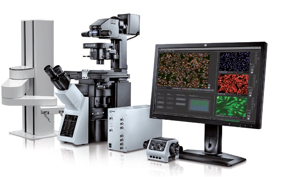
Olympus, Tokyo, has launched in the United States the ScanR high-content screening station, a cell-imaging solution that applies artificial intelligence (AI) to biological research. The unit combines the modularity and flexibility of a microscope-based setup with the automation, speed, throughput, and reproducibility of a high-content screening station.
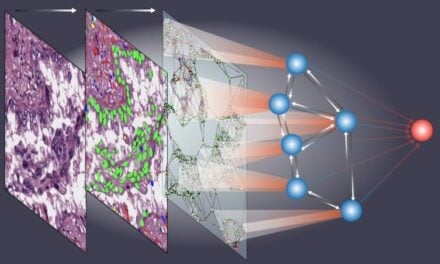
The ScanR station acquires pairs of images that are processed by the software to generate an image analysis model. Olympus’ optimized deep-learning technology, which is based on a dedicated convolutional neural network architecture, provides powerful and flexible learned analysis protocols. No human data annotations are required, which allows large numbers of examples to be used, to enable the full potential of the deep-learning technology.

Figure 1: Self-learning microscopy: AI prediction of nuclei positions (blue), green fluorescent protein (GFP) histone 2B labels showing nuclei (green), and raw brightfield transmission image (gray).
After a one-time training phase, ScanR AI enables the system to automatically analyze new data by incorporating the learned analysis protocol into its assay-based workflow. Because the user has full control in designing the training experiment, many challenging analysis conditions can be covered during the training phase. Subsequently, application of AI improves the accuracy and robustness of the system’s analysis and results.
The ScanR system performs fully automated image acquisition and analysis of multiwell plates, slides, and custom-built arrays. Throughout the automated process of image acquisition, the ScanR system maintains the focus plane using a combination of software algorithms and hardware, including TruFocus Z-drift compensation. Images are automatically analyzed during acquisition to minimize analysis time, and all units are precisely synchronized by a real-time controller to maximize the acquisition speed.
Olympus’ self-learning microscopy technology reduces photobleaching and improves acquisition speed, measurement sensitivity, and accuracy, to facilitate longer observations with reduced influence on cell viability.
For more information, visit Olympus Life Sciences.


