AI-Assisted Digital Parasitology Workflow Shows Strong Agreement with Traditional Microscopy
Peer-reviewed study found 98% concordance between Techcyte's digital workflow and brightfield microscopy in routine clinical laboratory testing.

Peer-reviewed study found 98% concordance between Techcyte's digital workflow and brightfield microscopy in routine clinical laboratory testing.

Researchers demonstrated that AI could accurately identify drug-resistant infections from microscopy images, reducing diagnostic time.


The CQ3000 has been designed to capture 3D microscopic images of live cell cultures in high definition at high speed.


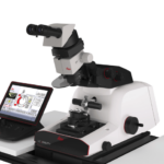
Nikon’s Eclipse Ni-L upright microscope features specially balanced LED illumination with properties similar to natural light for more accurate displays of the original color rendition of a specimen.
Read More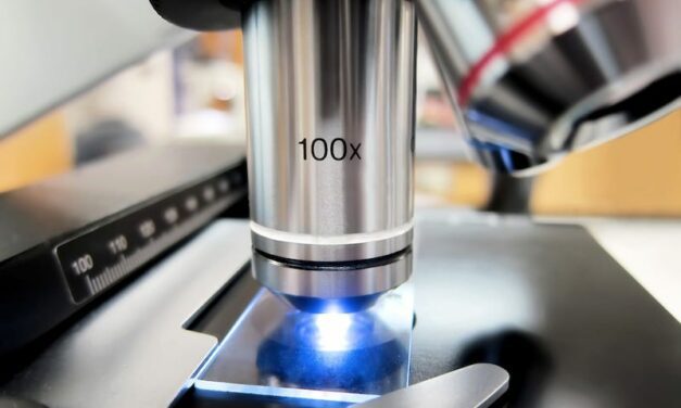
Children’s Colorado has announced its official Electron Microscopy (EM) accreditation to diagnose primary ciliary dyskinesia (PCD).
Read More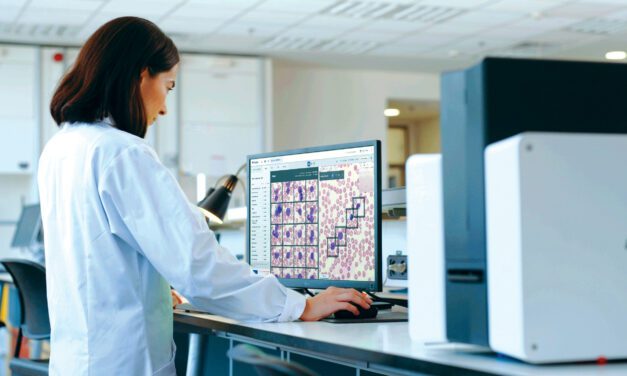
Scopio’s digital morphology and analysis system significantly reduced one medical center’s lab turnaround time for PBS review.
Read More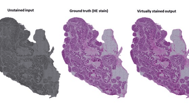
Researchers have developed an AI-based method for virtual staining of histopathological tissue samples that will replace chemical staining.
Read More
A team from the Max Planck Institute use artificial intelligence to evaluate surgical biopsy data, creating a kind of artificial pathologist.
Read More
Brillouin microscopy can map cell and tissue stiffness often associated with early signs of such diseases as cancer and Alzheimer’s.
Read More
The method leverages AI to improve the quality of images opening the possibility for real-time diagnoses during surgery.
Read More
A new, multiparameter approach may lead to liquid biopsies that will diagnose cancer with unprecedented accuracy.
Read More
One of the challenges of microscopy camera systems is that they don’t produce the same color rendering as the human eye.
Read More
Scopio Labs’ X100HT device with Peripheral Blood Smear (PBS) Application for hematological analysis has received U.S. FDA 510(k) clearance.
Read More
Nonlinear optical microscopy enables comprehensive & informative analysis of various biochemical phenomena despite a confined field of view.
Read More
Researchers in Korea have recently developed a new slow light technique to easily visualize viruses using an optical microscope.
Read More
Researchers have developed a microscopy system for non-invasive conjunctival goblet cells examination in patients.
Read More
Researchers has developed a technology that could replace conventional biopsies and histology with real-time imaging within the living body.
Read More
Learn how CLP’s editor discovered firsthand how clinical laboratories and laboratorians are being taken for granted.
Read More